Biomarkers for Anti-Angiogenic Therapy in Cancer

Abstract
:1. Introduction
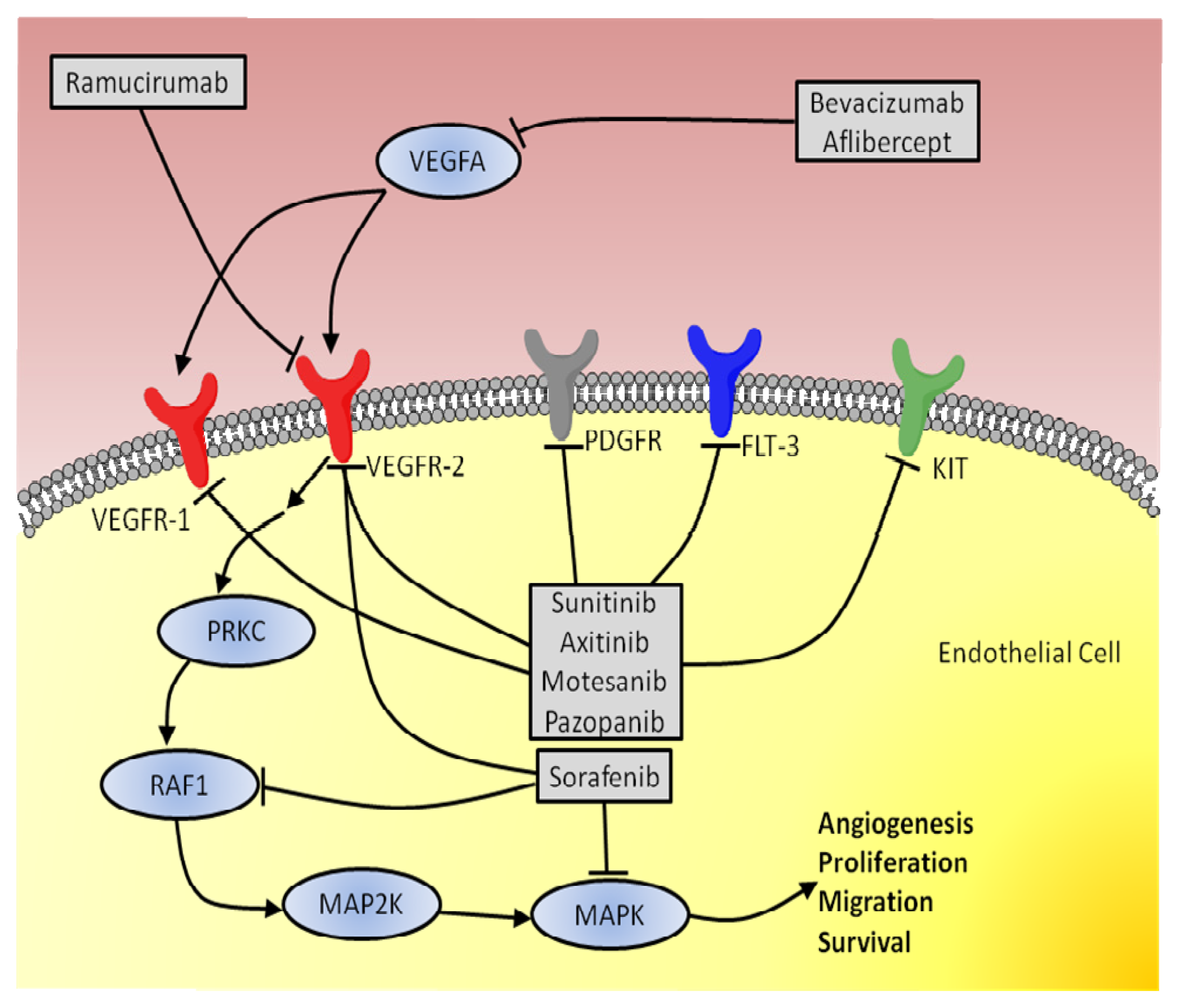
2. Anti-Angiogenic Therapy
3. Biomarkers
3.1. Biomarkers in Colorectal Cancer
3.1.1. Circulating Biomarkers
3.1.2. Genetic Biomarkers
3.1.3. Physiologic Biomarkers
3.2. Biomarkers in Breast Cancer
3.2.1. Circulating Biomarkers
3.2.2. Genetic Biomarkers
3.2.3. Physiologic Biomarkers
3.2.4. Tissue Biomarkers
3.3. Biomarkers in Thyroid Cancer
3.3.1. Circulating Biomarkers
3.3.2. Tissue Biomarkers
3.4. Biomarkers in Renal Cancer
3.4.1. Circulating Biomarkers
3.4.2. Genetic Biomarkers
Single Nucleotide Polymorphisms
3.5. Biomarkers in Prostate Cancer
3.6. Future Developments
4. Conclusions
Acknowledgements
Conflict of Interest
Abbreviations
| AMACR | Alpha-methylacyl-CoA racemase |
| Ang | Angiopoietin |
| ATP | Adenosine triphosphate |
| BRAF | Raf murine sarcoma viral oncogene homolog B1 |
| CAF | Cytokine and angiogenic factor |
| CAIX | Carbonic anhydrase IX (CAIX) |
| CEC | Circulating endothelial cells |
| CD | Cluster of differentiation |
| CR | Castration resistance |
| CT | Computer tomography |
| CTC | Circulating tumor cells |
| CXCR4 | CXC motif, Chemokine receptor type 4 |
| DNA | Deoxyribonucleic acid |
| DTC | Differentiated thyroid cancer |
| FDA | Food and Drug Administration |
| FDG | 18F-fluorodeoxyglucose |
| FGF | Fibroblast growth factor |
| FLT-3 | Fms-like tyrosine kinase 3 |
| Fluorouracil | Folinic acid |
| FOLFIRI | Irinotecan combination therapy |
| FOLFOX4 | Oxaliplatin combination therapy |
| HER2 | Human Epidermal Growth Factor Receptor 2 |
| HIF | Hypoxia-inducible transcription factor |
| ICAM | Intercellular adhesion molecule |
| IEF | Isoelectric focusing |
| IgG | Immunoglobulin G |
| IL | Interleukin |
| K-ras | Kirsten rat sarcoma viral oncogene homolog |
| KDR | Kinase insert domain receptor |
| KIT | Mast/stem cell growth factor receptor |
| LDH | Lactate dehydrogenase |
| MAPK | Mitogen-activated protein kinase |
| MAP2K | Mitogen-activated protein kinase kinase |
| mCRPC | Metastatic castration-resistant prostate cancer |
| MMP | Matrix metalloproteinase |
| MRI | Magnetic resonance imaging |
| MTC | Medullary thyroid carcinoma |
| NGAL | Neutrophil gelatinase associated lipocalin |
| NIH | National Institutes of Health |
| NO | Nitric oxide |
| OS | Overall survival |
| PC | Prostate cancer |
| PET | Positron emission tomography |
| PFS | Progression-free survival |
| PlGF | Placenta growth factor |
| PRKC | Protein kinase C |
| PSA | Prostate-specific antigen |
| PTC | Papillary thyroid cancer |
| PTTG | Pituitary Tumor-Transforming Gene 1-Interacting Protein |
| RAF1 | Proto-oncogene c-RAF |
| RCC | Renal cell carcinomas |
| RIBBON-1 | Regimens in Bevacizumab for Breast Oncology-1 |
| RNA | Ribonucleic acid |
| SCID | Severe combined immunodeficiency |
| SNP | Single Nucleotide Polymorphisms |
| TARGET | Treatment Approaches in Renal Cancer Global Evaluation Trial |
| Tg | Thyroglublin |
| TKI | Tyrosine kinase inhibitor |
| TNM | Tumor Node Metastasis system |
| TTP | Time to progression |
| ULN | Upper limit of normal |
| VCAM | Vascular cell adhesion molecule |
| VEGF | Vascular endothelial growth factors |
| VHL | Von Hippel-Lindau tumor suppressor |
| XELIRI | Capecitabine and Irinotecan combination therapy |
| XELOX | Capecitabine and Oxaliplatin combination therapy. |
References
- Straume, O.; Chappuis, P.O.; Salvesen, H.B.; Halvorsen, O.J.; Haukaas, S.A.; Goffin, J.R.; Bégin, L.R.; Foulkes, W.D.; Akslen, L.A. Prognostic importance of glomeruloid microvascular proliferation indicates an aggressive angiogenic phenotype in human cancers. Cancer Res 2002, 62, 6808–6811. [Google Scholar]
- Folkman, J. Tumor angiogenesis: Therapeutic implications. N. Engl. J. Med 1971, 285, 1182–1186. [Google Scholar]
- Arnold, F.; West, D.C. Angiogenesis in wound healing. Pharmacol. Ther 1991, 52, 407–422. [Google Scholar]
- Reynolds, L.P.; Redmer, D.A. Angiogenesis in the placenta. Biol. Reprod 2001, 64, 1033–1040. [Google Scholar]
- Folkman, J. Angiogenesis in cancer, vascular, rheumatoid and other disease. Nat. Med 1995, 1, 27–31. [Google Scholar]
- Hanahan, D.; Folkman, J. Patterns and emerging mechanisms of the angiogenic switch during tumorigenesis. Cell 1996, 86, 353–364. [Google Scholar]
- Skobe, M.; Rockwell, P.; Goldstein, N.; Vosseler, S.; Fusenig, N.E. Halting angiogenesis suppresses carcinoma cell invasion. Nat. Med 1997, 3, 1222–1227. [Google Scholar]
- Sullivan, R.; Graham, C.H. Hypoxia-driven selection of the metastatic phenotype. Cancer Metastasis. Rev 2007, 26, 319–331. [Google Scholar]
- Ferrara, N. Role of vascular endothelial growth factor. Am. J. Physiol. Cell Physiol 2001, 280, C1358–C1366. [Google Scholar]
- Ferrara, N.; Gerber, H.P.; LeCouter, J. The biology of VEGF and its receptors. Nat. Med 2003, 9, 666–669. [Google Scholar]
- Tammela, T.; Enholm, B.; Alitalo, K.; Paavonen, K. The biology of vascular endothelial growth factors. Cardiovasc. Res 2005, 65, 550–563. [Google Scholar]
- Kowanetz, M.; Ferrara, N. Vascular endothelial growth factor signaling pathways, therapeutic perspective. Clin. Cancer Res 2006, 12, 5018–5022. [Google Scholar]
- Holderfield, M.T.; Hughes, C.C. Crosstalk between vascular endothelial growth factor, notch, and transforming growth factor-beta in vascular morphogenesis. Circ. Res 2008, 102, 637–652. [Google Scholar]
- Bergers, G.; Hanahan, D. Modes of resistance to anti-angiogenic therapy. Nat. Rev. Cancer 2008, 8, 592–603. [Google Scholar]
- Carmeliet, P.; Ng, Y.S.; Nuyens, D.; Theilmeier, G.; Brusselmans, K.; Cornelissen, I.; Ehler, E.; Kakkar, V.V.; Stalmans, I.; Mattot, V.; et al. Impaired myocardial angiogenesis and ischemic cardiomyopathy in mice lacking the vascular endothelial growth factor isoforms VEGF(164) and VEGF(188). Nat. Med 1999, 5, 495–502. [Google Scholar]
- Valtola, R.; Salven, P.; Heikkilä, P.; Taipale, J.; Joensuu, H.; Rehn, M.; Pihlajaniemi, T.; Weich, H.; de Waal, R.; Alitalo, K. VEGFR-3 and its ligand VEGF-C are associated with angiogenesis in breast cancer. Am. J. Pathol 1999, 154, 1381–1390. [Google Scholar]
- Roskoski, R. Vascular endothelial growth factor (VEGF) signaling in tumor progression. Crit. Rev. Oncol. Hematol 2007, 62, 179–213. [Google Scholar]
- Ruohola, J.K.; Valve, E.M.; Karkkainen, M.J.; Joukov, V.; Alitalo, K.; Härkönen, P.L. Vascular endothelial growth factors are differentially regulated by steroid hormones and antiestrogens in breast cancer cells. Mol. Cell Endocrinol 1999, 149, 29–40. [Google Scholar]
- Dumont, N.; Arteaga, C.L. Transforming growth factor-beta and breast cancer—Tumor promoting effects of transforming growth factor-beta. Breast Cancer Res 2000, 2, 125–132. [Google Scholar]
- Gerber, H.P.; McMurtrey, A.; Kowalski, J.; Yan, M.; Keyt, B.A.; Dixit, V.; Ferrara, N. Vascular endothelial growth factor regulates endothelial cell survival through the phosphatidylinositol 3′-kinase/Akt signal transduction pathway. Requirement for Flk-1/KDR activation. Requirement for Flk-1/KDR activation. J. Biol. Chem 1998, 273, 30336–30343. [Google Scholar]
- Dor, Y.; Porat, R.; Keshet, E. Vascular endothelial growth factor and vascular adjustments to perturbations in oxygen homeostasis. Am. J. Physiol. Cell Physiol 2001, 280, C1367–C1374. [Google Scholar]
- Infanger, M.; Faramarzi, S.; Grosse, J.; Kurth, E.; Ulbrich, C.; Bauer, J.; Wehland, M.; Kreutz, R.; Kossmehl, P.; Paul, M.; et al. Expression of vascular endothelial growth factor and receptor tyrosine kinases in cardiac ischemia/reperfusion injury. Cardiovasc. Pathol. 2007, 16, 291–299. [Google Scholar]
- Semenza, G. Signal transduction to hypoxia-inducible factor 1. Biochem. Pharmacol 2002, 64, 993–998. [Google Scholar]
- Ferrara, N.; Davis-Smyth, T. The biology of vascular endothelial growth factor. Endocr. Rev 1997, 18, 4–25. [Google Scholar]
- Herbert, S.P.; Stainier, D.Y. Molecular control of endothelial cell behavior during blood vessel morphogenesis. Nat. Rev 2011, 12, 551–564. [Google Scholar]
- Shibuya, M.; Yamaguchi, S.; Yamane, A.; Ikeda, T.; Tojo, A.; Matsushime, H.; Sato, M. Nucleotide sequence and expression of a novel human receptor-type tyrosine kinase gene (flt) closely related to the fms family. Oncogene 1990, 5, 519–524. [Google Scholar]
- Terman, B.I.; Carrion, M.E.; Kovacs, E.; Rasmussen, B.A.; Eddy, R.L.; Shows, T.B. Identification of a new endothelial cell growth factor receptor tyrosine kinase. Oncogene 1991, 6, 1677–1683. [Google Scholar]
- Karkkainen, M.J.; Mäkinen, T.; Alitalo, K. Lymphatic endothelium, a new frontier of metastasis research. Nat. Cell Biol 2002, 4, E2–E5. [Google Scholar]
- Ferrara, N. Vascular endothelial growth factor, basic science and clinical progress. Endocr. Rev 2004, 25, 581–611. [Google Scholar]
- Alitalo, K.; Tammela, T.; Petrova, T.V. Lymphangiogenesis in development and human disease. Nature 2005, 438, 946–953. [Google Scholar]
- Paavonen, K.; Puolakkainen, P.; Jussila, L.; Jahkola, T.; Alitalo, K. Vascular endothelial growth factor receptor-3 in lymphangiogenesis in wound healing. Am. J. Pathol 2000, 156, 1499–1504. [Google Scholar]
- Tammela, T.; Zarkada, G.; Nurmi, H.; Jakobsson, L.; Heinolainen, K.; Tvorogov, D.; Zheng, W.; Franco, C.A.; Murtomäki, A.; Aranda, E.; et al. VEGFR-3 controls tip to stalk conversion at vessel fusion sites by reinforcing Notch signalling. Nat. Cell Biol 2011, 3, 1202–1213. [Google Scholar]
- Takahashi, H.; Shibuya, M. The vascular endothelial growth factor (VEGF)/VEGF receptor system and its role under physiological and pathological conditions. Clin. Sci 2005, 109, 227–241. [Google Scholar]
- Olsson, A.K.; Dimberg, A.; Kreuger, J.; Claesson-Welsh, L. VEGF receptor signaling—In control of vascular function. Nat. Rev. Mol. Cell Biol 2006, 7, 359–371. [Google Scholar]
- Park, J.E.; Chen, H.H.; Winer, J.; Houck, K.A.; Ferrara, N. Placenta growth factor. Potentiation of vascular endothelial growth factor bioactivity, in vitro and in vivo, and high affinity binding to Flt-1 but not to Flk-1/KDR. J. Biol. Chem 1994, 269, 25646–25654. [Google Scholar]
- Hiratsuka, S.; Minowa, O.; Kuno, J.; Noda, T.; Shibuya, M. Flt-1 lacking the tyrosine kinase domain is sufficient for normal development and angiogenesis in mice. Proc. Natl. Acad. Sci. USA 1998, 4, 9349–9354. [Google Scholar]
- Infanger, M.; Schmidt, O.; Kossmehl, P.; Grad, S.; Ertel, W.; Grimm, D. Vascular endothelial growth factor serum level is strongly enhanced after burn injury and correlated with local and general tissue edema. Burns 2004, 30, 305–311. [Google Scholar]
- Infanger, M.; Shakibaei, M.; Kossmehl, P.; Hollenberg, S.M.; Grosse, J.; Faramarzi, S.; Schulze-Tanzil, G.; Paul, M.; Grimm, D. Intraluminal application of vascular endothelial growth factor enhances healing of microvascular anastomosis in a rat model. J. Vasc. Res 2005, 42, 202–213. [Google Scholar]
- Infanger, M.; Grosse, J.; Westphal, K.; Leder, A.; Ulbrich, C.; Paul, M.; Grimm, D. Vascular Endothelial Growth Factor induces extracellular matrix proteins and osteopontin in the umbilical artery. Ann. Vasc. Surg 2008, 22, 273–284. [Google Scholar]
- Alon, T.; Hemo, I.; Itin, A.; Pe’er, J.; Stone, J.; Keshet, E. Vascular endothelial growth factor acts as a survival factor for newly formed retinal vessels and has implications for retinopathy of prematurity. Nat. Med 1995, 1, 1024–1028. [Google Scholar]
- Gerber, H.P.; Dixit, V.; Ferrara, N. Vascular Endothelial Growth Factor induces expression of the antiapoptotic proteins Bcl-2 and A1 in vascular endothelial cells. J. Biol. Chem 1998, 273, 13313–13316. [Google Scholar]
- Benjamin, L.E.; Golijanin, D.; Itin, A.; Pode, D.; Keshet, E. Selective ablation of immature blood vessels in established human tumors follows vascular endothelial growth factor withdrawal. J. Clin. Invest 1999, 103, 159–165. [Google Scholar]
- Infanger, M.; Kossmehl, P.; Shakibaei, M.; Baatout, S.; Witzing, A.; Grosse, J.; Bauer, J.; Cogoli, A.; Faramarzi, S.; Derradji, H.; et al. Induction of three-dimensional assembly and increase in apoptosis of human endothelial cells by simulated microgravity. Impact of vascular endothelial growth factor. Apoptosis 2006, 11, 749–764. [Google Scholar]
- Ferrara, N.; Hillan, K.; Gerber, H.P.; Novotny, W. Discovery and development of Bevacizumab, an anti VEGF antibody for treating cancer. Nat. Rev. Drug Discov 2004, 3, 391–398. [Google Scholar]
- Reck, M.; von Pawel, J.; Zatloukal, P.; Ramlau, R.; Gorbounova, V.; Hirsh, V.; Leighl, N.; Mezger, J.; Archer, V.; Moore, N.; et al. BO17704 Study Group. Overall survival with cisplatin-gemcitabina and Bevacizumab or placebo as first line therapy for non-squamous NSCLC: Results from a randomized phase III trial (AVAiL). Ann. Oncol 2010, 21, 1804–1809. [Google Scholar]
- Sandler, A.; Gray, R.; Perry, M.C.; Brahmer, J.; Schiller, J.H.; Dowlati, A.; Lilenbaum, R.; Johnson, D.H. Paclitaxel-Carboplatin alone or with Bevacizumab for non-small-cell lung cancer. N. Engl. J. Med 2006, 355, 2542–2550. [Google Scholar]
- Johnson, D.H.; Fehrenbacher, L.; Novotny, W.F.; Herbst, R.S.; Nemunaitis, J.J.; Jablons, D.M.; Langer, C.J.; DeVore, R.F., 3rd; Gaudreault, J.; Damico, L.A.; et al. Randomized phase II trial comparing Bevacizumab plus Carboplatin and Paclitaxel with Carboplatin and Paclitaxel alone in previously untreated locally advanced or metastatic non-small-cell lung cancer. J. Clin. Oncol 2004, 22, 2184–2191. [Google Scholar]
- Herbst, R.S.; O’Neill, V.J.; Fehrenbacher, L.; Belani, C.P.; Bonomi, P.D.; Hart, L.; Melnyk, O.; Ramies, D.; Lin, M.; Sandler, A. Phase II study of efficacy and safety of Bevacizumab in combination with chemotherapy or Erlotinib compared with chemotherapy alone for treatment of recurrent or refractory non-small-cell lung cancer. J. Clin. Oncol 2007, 25, 4743–4750. [Google Scholar]
- Miller, K.D.; Chap, L.I.; Holmes, F.A.; Cobleigh, M.A.; Marcom, P.K.; Fehrenbacher, L.; Dickler, M.; Overmoyer, B.A.; Reimann, J.D.; Sing, A.P.; et al. Randomized phase III trial of Capecitabine compared with Bevacizumab plus Capecitabine in patient with previously treated metastatic breast cancer. J. Clin. Oncol 2005, 23, 792–799. [Google Scholar]
- Miller, K.; Wang, M.; Gralow, J.; Dickler, M.; Cobleigh, M.; Perez, E.A.; Shenkier, T.; Cella, D.; Davidson, N.E. Paclitaxel with Bevacizumab versus Paclitaxel alone in metastatic breast cancer. N. Engl. J. Med 2007, 357, 2666–2676. [Google Scholar]
- Robert, N.J.; Dieras, V.; Glaspy, J.; Brufsky, A.; Bondarenko, I.; Lipatov, O.; Perez, E.; Yardley, D.; Zhou, X.; Phan, S. RIBBON-1, randomized, double blind, placebo controlled phase III trial of chemotherapy with or without Bevacizumab for first line treatment of HER2 negative locally recurrent or metastatic breast cancer. ASCO Present. J. Clin. Oncol 2009, 27, 15s. [Google Scholar]
- Miles, D.W.; Chan, A.; Dirix, L.Y.; Cortés, J.; Pivot, X.; Tomczak, P.; Delozier, T.; Sohn, J.H.; Provencher, L.; Puglisi, F.; et al. Phase III study of Bevacizumab plus Docetaxel campared with placebo plus docetaxel for the first line treatment of human epidermal growth factor receptor-2-negative metastatic breast cancer. J. Clin. Oncol 2010, 28, 3239–3247. [Google Scholar]
- Brufsky, A.M.; Hurvitz, S.; Perez, E.; Swamy, R.; Valero, V.; O’Neill, V.; Rugo, H.S. RIBBON-2, a randomized, double-blind, placebo-controlled, phase III trial evaluating the efficacy and safety of Bevacizumab in combination with chemotherapy for second-line treatment of human epidermal growth factor receptor 2-negative metastatic breast cancer. J. Clin. Oncol 2011, 29, 4286–4293. [Google Scholar]
- Kabbinavar, F.; Hurwitz, H.I.; Fehrenbacher, L.; Meropol, N.J.; Novotny, W.F.; Lieberman, G.; Griffing, S.; Bergsland, E. Phase II randomized trial comparing Bevacizumab plus Fluorouracil(FU)/Leucovorin(LV) with FU/LV alone in patients with metastatic colorectal cancer. J. Clin. Oncol 2003, 21, 60–65. [Google Scholar]
- Kabbinavar, F.F.; Schulz, J.; McCleod, M.; Patel, T.; Hamm, J.T.; Hecht, J.R.; Mass, R.; Perrou, B.; Nelson, B.; Novotny, W.F. Addition of Bevacizumab to bolus Fluorouracil and Leucovorin in first line metastatic colorectal cancer, results of randomized phase II trial. J. Clin. Oncol 2005, 23, 3697–3705. [Google Scholar]
- Hurwitz, H.; Fehrenbacher, L.; Novotny, W.; Cartwright, T.; Hainsworth, J.; Heim, W.; Berlin, J.; Baron, A.; Griffing, S.; Holmgren, E.; et al. Bevacizumab plus Irinotecan, Flouorouracil and Leucovorin for metastatic colorectal cancer. N. Engl. J. Med 2004, 350, 2335–2342. [Google Scholar]
- Saltz, L.B.; Clarke, S.; Díaz-Rubio, E.; Scheithauer, W.; Figer, A.; Wong, R.; Koski, S.; Lichinitser, M.; Yang, T.S.; Rivera, F.; et al. Bevacizumab in combination with oxaliplatin based chemotherapy as first line therapy in metastatic colorectal cancer: A randomized phase III study. J. Clin. Oncol 2008, 26, 2013–2019. [Google Scholar]
- Tebbutt, N.C.; Wilson, K.; Gebski, V.J.; Cummins, M.M.; Zannino, D.; van Hazel, G.A.; Robinson, B.; Broad, A.; Ganju, V.; Ackland, S.P.; et al. Capecitabine, Bevacizumab and Mitomycin in first line treatment of metastatic colorectal cancer: Results of the Australian gastrointestinal trials group randomized phase III MAX study. J. Clin. Oncol 2010, 28, 3191–3198. [Google Scholar]
- Rini, B.I.; Halabi, S.; Rosenberg, J.E.; Stadler, W.M.; Vaena, D.A.; Archer, L.; Atkins, J.N.; Picus, J.; Czaykowski, P.; Dutcher, J.; et al. Phase III trial of Bevacizumab plus interferon alfa versus interferon alfa monotherapy in patients with metastatic renal cell carcinoma: Final results of CALGB 90206. J. Clin. Oncol 2010, 28, 1–7. [Google Scholar]
- Escudier, B.; Pluzanska, A.; Koralewski, P.; Ravaud, A.; Bracarda, S.; Szczylik, C.; Chevreau, C.; Filipek, M.; Melichar, B.; Bajetta, E.; et al. AVOREN Trial investigators. Bevacizumab plus interferon alfa-2a for treatment of metastatic renal cell carcinoma: A randomized double blind phase III trial. Lancet 2007, 370, 2103–2111. [Google Scholar]
- Ohtsu, A.; Shah, M.A.; van Cutsem, E.; Rha, S.Y.; Sawaki, A.; Park, S.R.; Lim, H.Y.; Yamada, Y.; Wu, J.; Langer, B.; et al. Bevacizumab in combination with chemotherapy as first-line therapy in advanced gastric cancer: A randomized, double-blind, placebo-controlled phase III study. J. Clin. Oncol 2011, 29, 3968–3976. [Google Scholar]
- Kindler, H.L.; Niedzwiecki, D.; Hollis, D.; Sutherland, S.; Schrag, D.; Hurwitz, H.; Innocenti, F.; Mulcahy, M.F.; O’Reilly, E.; Wozniak, T.F.; et al. Gemcitabine plus Bevacizumab compared with Gemcitabine plus placebo in patients with advanced pancreatic cancer: Phase III trial of the cancer and leukemia group B (CALGB 80303). J. Clin. Oncol 2010, 28, 3617. [Google Scholar]
- Van Cutsem, E.; Vervenne, W.L.; Bennouna, J.; Humblet, Y.; Gill, S.; van Laethem, J.L.; Verslype, C.; Scheithauer, W.; Shang, A.; Cosaert, J.; et al. Phase III trial of Bevacizumab in combination with Gemcitabine and Erlotinib in patients with metastatic pancreatic cancer. J. Clin. Oncol 2009, 27, 2231–2237. [Google Scholar]
- Kelly, W.K.; Halabi, S.; Carducci, M.; George, D.; Mahoney, J.F.; Stadler, W.M.; Morris, M.; Kantoff, P.; Monk, J.P.; Kaplan, E.; et al. Randomized, double-blind, placebo-controlled phase III trial comparing docetaxel and prednisone with or without bevacizumab in men with metastatic castration-resistant prostate cancer: CALGB 90401. J. Clin. Oncol 2012, 30, 1534–1540. [Google Scholar]
- Kim, K.B.; Sosman, J.A.; Fruehauf, J.P.; Linette, G.P.; Markovic, S.N.; McDermott, D.F.; Weber, J.S.; Nguyen, H.; Cheverton, P.; Chen, D.; et al. BEAM: A randomized phase II study evaluating the activity of Bevacizumab in combination with Carboplatinum plus Paclitaxel in patients with previously untreated advanced melanoma. J. Clin. Oncol 2012, 30, 34–41. [Google Scholar]
- Tew, W.P.; Colombo, N.; Ray-Coquard, I.; Oza, A.; del Campo, J.; Scambia, G.; Spriggs, D. VEGF-Trap for patients (pts) with recurrent platinum-resistant epithelial ovarian cancer (EOC), preliminary results of a randomized, multicenter phase II study. ASCO Meet. Abstr 2007, 25, 5508. [Google Scholar]
- Tang, P.; Cohen, S.J.; Bjarnason, G.A.; Kollmannsberger, C.; Virik, K.; MacKenzie, M.J.; Brown, J.; Wang, L.; Chen, A.P.; Moore, M.J. Phase II trial of aflibercept (VEGF Trap) in previously treated patients with metastatic colorectal cancer (MCRC): A PMH phase II consortium trial. ASCO Meet. Abstr 2008, 26, 4027. [Google Scholar]
- Massarelli, E.; Miller, V.A.; Leighl, N.B.; Rosen, P.J.; Albain, K.S.; Hart, L.L.; Melnyk, O.; Sternas, L.; Ackerman, J.; Herbst, R.S. Phase II study of the efficacy and safety of intravenous (IV) AVE0005 (VEGF Trap) given every 2 weeks in patients (Pts) with platinum- and erlotinib-resistant adenocarcinoma of the lung (NSCLA). ASCO Meet. Abstr 2007, 25, 7627. [Google Scholar]
- Townsley, C.; Hirte, H.; Hoskins, P.; Buckanovich, R.; Mackay, H.; Welch, S.; Wang, L.; Polintan, R.; Chen, A.; Oza, A.M. A phase II study of aflibercept (VEGF trap) in recurrent or metastatic gynecologic soft-tissue sarcomas: A study of the Princess Margaret Hospital Phase II Consortium. ASCO Meet. Abstr 2009, 27, 5591. [Google Scholar]
- Twardowski, P.; Stadler, W.M.; Frankel, P.; Lara, P.N.; Ruel, C.; Chatta, G.; Heath, E.I.; Quinn, D.I.; Gandara, D.R. Phase II study of aflibercept (VEGFTrap) in patients with recurrent or metastatic transitional cell carcinoma (TCC) of the urothelium: A California Cancer Consortium trial. ASCO Meet. Abstr 2009, 27, e16030. [Google Scholar]
- Tarhini, A.A.; Christensen, S.; Frankel, P.; Margolin, K.; Ruel, C.; Shipe-Spotloe, J.; DeMark, M.; Kirkwood, J.M. Phase II study of aflibercept (VEGF trap) in recurrent inoperable stage III or stage IV melanoma of cutaneous or ocular origin. ASCO Meet. Abstr 2009, 27, 9028. [Google Scholar]
- De Groot, J.F.; Wen, P.Y.; Lamborn, K.; Chang, S.; Cloughesy, T.F.; Chen, A.P.; DeAngelis, L.M.; Mehta, M.P.; Gilbert, M.R.; Yung, W.K.; et al. Phase II single arm trial of aflibercept in patients with recurrent temozolomide-resistant glioblastoma: NABTC 0601. ASCO Meet. Abstr 2008, 26, 2020. [Google Scholar]
- Lu, D.; Jimenez, X.; Zhang, H.; Bohlen, P.; Witte, L.; Zhu, Z. Selection of high affinity human neutralizing antibodies to VEGFR2 from a large antibody phage display library for antiangiogenesis therapy. Int. J. Cancer 2002, 97, 393–399. [Google Scholar]
- Spratlin, J. Ramucirumab (IMC-1121B), Monoclonal antibody inhibition of vascular endothelial growth factor receptor-2. Curr. Oncol. Rep 2011, 13, 97–102. [Google Scholar]
- Wood, L. Sunitinib malate for the treatment of renal cell carcinoma. Expert Opin. Pharmacother 2012, 13, 1323–1336. [Google Scholar]
- Sekkate, S.; Kairouani, M.; Abahssain, H.; Serji, B.; Boutayeb, S.; Mrabti, H.; Errihani, H. Gastrointestinal stromal tumors. Presse. Med 2012, 41, 917–926. [Google Scholar]
- Escudier, B.; Eisen, T.; Stadler, W.M.; Szczylik, C.; Oudard, S.; Staehler, M.; Negrier, S.; Chevreau, C.; Desai, A.A.; Rolland, F.; et al. Sorafenib for treatment of renal cell carcinoma, Final efficacy and safety results of the phase III treatment approaches in renal cancer global evaluation trial. J. Clin. Oncol 2009, 27, 3312–3318. [Google Scholar]
- Zhu, A.X. Development of sorafenib and other molecularly targeted agents in hepatocellular carcinoma. Cancer 2008, 112, 250–259. [Google Scholar]
- Scagliotti, G.V.; Vynnychenko, I.; Park, K.; Ichinose, Y.; Kubota, K.; Blackhall, F.; Pirker, R.; Galiulin, R.; Ciuleanu, T.E.; Sydorenko, O.; et al. International, randomized, placebo-controlled, double-blind phase III study of motesanib plus carboplatin/paclitaxel in patients with advanced nonsquamous non-small-cell lung cancer: MONET1. J. Clin. Oncol 2012, 30, 2829–2836. [Google Scholar]
- Coxon, A.; Bready, J.; Kaufman, S.; Estrada, J.; Osgood, T.; Canon, J.; Wang, L.; Radinsky, R.; Kendall, R.; Hughes, P.; et al. Anti-tumor activity of motesanib in a medullary thyroid cancer model. J. Endocrinol. Invest 2012, 35, 181–190. [Google Scholar]
- Gennigens, C.; Jerusalem, G. Pazopanib (Votrient) in the management of renal cell cancer and soft tissue sarcomas. Rev. Med. Liege 2012, 67, 437–442. [Google Scholar]
- Grimm, D.; Wise, P.; Lebert, M.; Richter, P.; Baatout, S. How and why does the proteome respond to microgravity? Expert Rev. Proteomics 2011, 8, 13–27. [Google Scholar]
- Pietsch, J.; Bauer, J.; Egli, M.; Infanger, M.; Wise, P.; Ulbrich, C.; Grimm, D. The effects of weightlessness on the human organism and mammalian cells. Curr. Mol. Med 2011, 11, 350–364. [Google Scholar]
- Grimm, D.; Infanger, M.; Westphal, K.; Ulbrich, C.; Pietsch, J.; Kossmehl, P.; Vadrucci, S.; Baatout, S.; Flick, B.; Paul, M.; et al. A delayed type of three-dimensional growth of human endothelial cells under simulated weightlessness. Tissue Eng. Part. A 2009, 15, 2267–2275. [Google Scholar]
- Grimm, D.; Bauer, J.; Ulbrich, C.; Westphal, K.; Wehland, M.; Infanger, M.; Aleshcheva, G.; Pietsch, J.; Ghardi, M.; Beck, M.; et al. Different responsiveness of endothelial cells to vascular endothelial growth factor and basic fibroblast growth factor added to culture media under gravity and simulated microgravity. Tissue Eng. Part A 2010, 16, 1559–1573. [Google Scholar]
- De Gruttola, V.G.; Clax, P.; DeMets, D.L.; Downing, G.J.; Ellenberg, S.S.; Friedman, L.; Gail, M.H.; Prentice, R.; Wittes, J.; Zeger, S.L. Considerations in the evaluation of surrogate endpoints in clinical trials. Summary of a National Institutes of Health workshop. Control. Clin. Trials 2001, 22, 485–502. [Google Scholar]
- Oldenhuis, C.N.; Oosting, S.F.; Gietema, J.A.; de Vries, E.G. Prognostic versus predictive value of biomarkers in oncology. Eur. J. Cancer 2008, 44, 946–953. [Google Scholar]
- McShane, L.M.; Altman, D.G.; Sauerbrei, W.; Taube, S.E.; Gion, M.; Clark, G.M. Statistics Subcommittee of NCI-EORTC Working Group on Cancer Diagnostics. Reporting recommendations for tumor MARKer prognostic studies (REMARK). Breast Cancer Res. Treat 2006, 100, 229–235. [Google Scholar]
- Ferlay, J.; Shin, H.R.; Bray, F.; Forman, D.; Mathers, C.; Parkin, D.M. GLOBOCAN 2008 v1.2, Cancer Incidence and Mortality Worldwide, IARC CancerBase No. 10 [Internet]; International Agency for Research on Cancer: Lyon, France, 2010. Available online: http://globocan.iarc.fr (accessed on 15 February 2013).
- Ince, W.L.; Jubb, A.M.; Holden, S.N.; Holmgren, E.B.; Tobin, P.; Sridhar, M.; Hurwitz, H.I.; Kabbinavar, F.; Novotny, W.F.; Hillan, K.J.; et al. Association of k-ras, b-raf, and p53 status with the treatment effect of Bevacizumab. J. Natl. Cancer Inst 2005, 97, 981–989. [Google Scholar]
- Jubb, A.M.; Hurwitz, H.I.; Bai, W.; Holmgren, E.B.; Tobin, P.; Guerrero, A.S.; Kabbinavar, F.; Holden, S.N.; Novotny, W.F.; Frantz, G.D.; et al. Impact of vascular endothelial growth factor-A expression, thrombospondin-2 expression, and microvessel density on the treatment effect of Bevacizumab in metastatic colorectal cancer. J. Clin. Oncol 2006, 24, 217–227. [Google Scholar]
- Cetin, B.; Kaplan, M.A.; Berk, V.; Ozturk, S.C.; Benekli, M.; Isıkdogan, A.; Ozkan, M.; Coskun, U.; Buyukberber, S. Prognostic factors for overall survival in patients with metastatic colorectal carcinoma treated with vascular endothelial growth factor-targeting agents. Asian Pac. J. Cancer Prev 2012, 13, 1059–1063. [Google Scholar]
- Kopetz, S.; Hoff, P.M.; Morris, J.S.; Wolff, R.A.; Eng, C.; Glover, K.Y.; Adinin, R.; Overman, M.J.; Valero, V.; Wen, S.; et al. Phase II trial of infusional fluorouracil, Irinotecan, and Bevacizumab for metastatic colorectal cancer, efficacy and circulating angiogenic biomarkers associated with therapeutic resistance. J. Clin. Oncol 2010, 28, 453–459. [Google Scholar]
- Goede, V.; Coutelle, O.; Neuneier, J.; Reinacher-Schick, A.; Schnell, R.; Koslowsky, T.C.; Weihrauch, M.R.; Cremer, B.; Kashkar, H.; Odenthal, M.; et al. Identification of serum angiopoietin-2 as a biomarker for clinical outcome of colorectal cancer patients treated with Bevacizumab-containing therapy. Br. J. Cancer 2010, 103, 1407–1414. [Google Scholar]
- Matsusaka, S.; Suenaga, M.; Mishima, Y.; Takagi, K.; Terui, Y.; Mizunuma, N.; Hatake, K. Circulating endothelial cells predict for response to Bevacizumab-based chemotherapy in metastatic colorectal cancer. Cancer Chemother. Pharmacol 2011, 68, 763–768. [Google Scholar]
- Matsusaka, S.; Mishima, Y.; Suenaga, M.; Terui, Y.; Kuniyoshi, R.; Mizunuma, N.; Hatake, K. Circulating endothelial progenitors and CXCR4-positive circulating endothelial cells are predictive markers for Bevacizumab. Cancer 2011, 117, 4026–4032. [Google Scholar]
- Ronzoni, M.; Manzoni, M.; Mariucci, S.; Loupakis, F.; Brugnatelli, S.; Bencardino, K.; Rovati, B.; Tinelli, C.; Falcone, A.; Villa, E.; et al. Circulating endothelial cells and endothelial progenitors as predictive markers of clinical response to Bevacizumab-based first-line treatment in advanced colorectal cancer patients. Ann. Oncol 2010, 21, 2382–2389. [Google Scholar]
- Manzoni, M.; Mariucci, S.; Delfanti, S.; Rovati, B.; Ronzoni, M.; Loupakis, F.; Brugnatelli, S.; Tinelli, C.; Villa, E.; Falcone, A.; et al. Circulating endothelial cells and their apoptotic fraction are mutually independent predictive biomarkers in Bevacizumab-based treatment for advanced colorectal cancer. J. Cancer Res. Clin. Oncol 2012, 138, 1187–1196. [Google Scholar]
- Singh, H.; Pohl, A.; El-Khoueiry, A.; Lurje, G.; Zhang, W.; Yang, D.; Ning, Y.; Shriki, J.; Iqbal, S.; Lenz, H. Use of genetic variants to predict clinical outcome in patients (pts) with metastatic colorectal cancer (mCRC) treated with first-line 5-FU or capecitabine in combination with oxaliplatin and Bevacizumab (FOLFOX/BV or XELOX/BV). J. Clin. Oncol 2009, 27, 15s. [Google Scholar]
- Hansen, T.F.; Christensen, R.D.; Andersen, R.F.; Garm Spindler, K.L.; Johnsson, A.; Jakobsen, A. The predictive value of single nucleotide polymorphisms in the VEGF system to the efficacy of first-line treatment with Bevacizumab plus chemotherapy in patients with metastatic colorectal cancer: Results from the Nordic ACT trial. Int. J. Colorectal Dis 2012, 27, 715–720. [Google Scholar]
- O’Brien, C.A.; Pollett, A.; Gallinger, S.; Dick, J.E. A human colon cancer cell capable of initiating tumour growth in immunodeficient mice. Nature 2007, 445, 106–110. [Google Scholar]
- Ricci-Vitiani, L.; Lombardi, D.G.; Pilozzi, E.; Biffoni, M.; Todaro, M.; Peschle, C.; De Maria, R. Identification and expansion of human colon-cancer-initiating cells. Nature 2007, 445, 111–115. [Google Scholar]
- Pohl, A.; Zhang, W.; Yang, D.; Lurje, G.; Ning, Y.; Khambata-Ford, S.; Langer, C.; Kahn, M.; Teo, J.L.; Lenz, H.J. Association of CD133 polymorphisms and clinical outcome in metastatic colorectal cancer (mCRC) patients (pts) treated with either first-line 5-FU + Bevacizumab (BV) or second-line Irinotecan (IR)/Cetuximab (CB) or IR alone. J. Clin. Oncol 2009, 27, 15s. [Google Scholar]
- Hurwitz, H.I.; Douglas, P.S.; Middleton, J.P.; Sledge, G.W.; Johnson, D.H.; Reardon, D.A.; Chen, D.; Rosen, O. Analysis of early hypertension and clinical outcome with bevacizumab: results from seven phase iii studies. Oncologist 2013, 18, 273–280. [Google Scholar]
- Jubb, A.M.; Harris, A.L. Biomarkers to predict the clinical efficacy of Bevacizumab in cancer. Lancet Oncol 2010, 11, 1172–1183. [Google Scholar]
- Scartozzi, M.; Galizia, E.; Chiorrini, S.; Giampieri, R.; Berardi, R.; Pierantoni, C.; Cascinu, S. Arterial hypertension correlates with clinical outcome in colorectal cancer patients treated with first-line Bevacizumab. Ann. Oncol 2009, 20, 227–230. [Google Scholar]
- Österlund, P.; Soveri, L.M.; Isoniemi, H.; Poussa, T.; Alanko, T.; Bono, P. Hypertension and overall survival in metastatic colorectal cancer patients treated with Bevacizumab-containing chemotherapy. Br. J. Cancer 2011, 104, 599–604. [Google Scholar]
- De Stefano, A.; Carlomagno, C.; Pepe, S.; Bianco, R.; de Placido, S. Bevacizumab-related arterial hypertension as a predictive marker in metastatic colorectal cancer patients. Cancer Chemother. Pharmacol 2011, 68, 1207–1213. [Google Scholar]
- Wehland, M.; Bauer, J.; Infanger, M.; Grimm, D. Target-based anti-angiogenic therapy in breast cancer. Curr. Pharm. Des 2012, 18, 4244–4257. [Google Scholar]
- Burstein, H.J.; Chen, Y.H.; Parker, L.M.; Savoie, J.; Younger, J.; Kuter, I.; Ryan, P.D.; Garber, J.E.; Chen, H.; Campos, S.M.; et al. VEGF as a marker for outcome among advanced breast cancer patients receiving anti-VEGF therapy with Bevacizumab and vinorelbine chemotherapy. Clin. Cancer Res 2008, 14, 7871–7877. [Google Scholar]
- Baar, J.; Silverman, P.; Lyons, J.; Fu, P.; Abdul-Karim, F.; Ziats, N.; Wasman, J.; Hartman, P.; Jesberger, J.; Dumadag, L.; et al. A vasculature-targeting regimen of preoperative docetaxel with or without Bevacizumab for locally advanced breast cancer: Impact on angiogenic biomarkers. Clin. Cancer Res 2009, 15, 3583–3590. [Google Scholar]
- Burstein, H.J.; Elias, A.D.; Rugo, H.S.; Cobleigh, M.A.; Wolff, A.C.; Eisenberg, P.D.; Lehman, M.; Adams, B.J.; Bello, C.L.; DePrimo, S.E.; et al. Phase II study of Sunitinib malate, an oral multitargeted tyrosine kinase inhibitor, in patients with metastatic breast cancer previously treated with an Anthracycline and a Taxane. J. Clin. Oncol 2008, 26, 1810–1816. [Google Scholar]
- Calleri, A.; Bono, A.; Bagnardi, A.; Quarna, J.; Mancuso, P.; Rabascio, C.; Dellapasqua, S.; Campagnoli, E.; Shaked, Y.; Goldhirsch, A.; et al. Predictive potential of angiogenic growth factors and circulating endothelial cells in breast cancer patients receiving metronomic chemotherapy plus Bevacizumab. Clin. Cancer Res 2009, 15, 7652–7657. [Google Scholar]
- Schneider, B.P.; Wang, M.; Radovich, M.; Sledge, G.W.; Badve, S.; Thor, A.; Flockhart, D.A.; Hancock, B.; Davidson, N.; Gralow, J.; et al. ECOG 2100. Association of vascular endothelial growth factor and vascular endothelial growth factor receptor-2 genetic polymorphisms with outcome in a trial of paclitaxel compared with paclitaxel plus Bevacizumab in advanced breast cancer: ECOG 2100. J. Clin. Oncol 2008, 26, 4672–4678. [Google Scholar]
- Hurwitz, H.; Douglas, P.S.; Middleton, J.P.W.; Sledge, G.; Johnson, D.H.; Reardon, D.A.; Chen, D.; Rosen, O. Analysis of early hypertension (HTN) and clinical outcome with Bevacizumab (BV). J. Clin. Oncol 2010, 28, 15s. [Google Scholar]
- Fountzilas, G.; Kourea, H.P.; Bobos, M.; Televantou, D.; Kotoula, V.; Papadimitriou, C.; Papazisis, K.T.; Timotheadou, E.; Efstratiou, I.; Koutras, A.; et al. Paclitaxel and Bevacizumab as first line combined treatment in patients with metastatic breast cancer, the Hellenic Cooperative Oncology Group experience with biological marker evaluation. Anticancer Res 2011, 31, 3007–3018. [Google Scholar]
- Busnardo, B.; de Vido, D. The epidemiology and etiology of differentiated thyroid carcinoma. Biomed. Pharmacother 2000, 54, 322–326. [Google Scholar]
- Okuieff, P.; Chen, Y.; Maguire, D.J.; Huser, A.K. Molecular markers of radiation-related normal tissue toxicity. Cancer Metastasis Rev 2008, 27, 363–374. [Google Scholar]
- Gilliland, F.D.; Hunt, W.C.; Morris, D.M.; Key, C.R. Prognostic factors for thyroid carcinoma. A population-based study of 15,698 cases from the Surveillance, Epidemiology and End Results (SEER) program 1973–1991. Cancer 1997, 79, 564–573. [Google Scholar]
- Durante, C.; Haddy, N.; Baudin, E.; Leboulleux, S.; Hartl, D.; Travagli, J.P.; Caillou, B.; Ricard, M.; Lumbroso, J.D.; de Vathaire, F.; et al. Long-term outcome of 444 patients with distant metastases from papillary and follicular thyroid carcinoma, benefits and limits of radioiodine therapy. J. Clin. Endocrinol. Metab 2006, 91, 2892–2899. [Google Scholar]
- Ain, K.B.; Lee, C.; Williams, K.D. Phase II trial of thalidomide for therapy of radioiodine-unresponsive and rapidly progressive thyroid carcinomas. Thyroid 2007, 17, 663–670. [Google Scholar]
- Schönberger, J.; Bauer, J.; Spruss, T.; Weber, G.; Chahoud, I.; Eilles, C.; Grimm, D. Establishment and characterization of the follicular thyroid carcinoma cell line ML-1. J. Mol. Med 2000, 78, 102–110. [Google Scholar]
- Schoenberger, J.; Grimm, D.; Kossmehl, P.; Infanger, M.; Kurth, E.; Eilles, C. Effects of PTK787/ZK222584, a tyrosine kinase inhibitor, on the growth of a poorly differentiated thyroid carcinoma, an animal study. Endocrinology 2004, 145, 1031–1038. [Google Scholar]
- Niedzwiecki, S.; Stepien, T.; Kopec, K.; Kuzdak, K.; Komorowski, J.; Krupinski, R.; Stepien, H. Angiopoietin 1 (Ang-1), angiopoietin 2 (Ang-2) and Tie-2 (a receptor tyrosine kinase) concentrations in peripheral blood of patients with thyroid cancers. Cytokine 2006, 36, 291–295. [Google Scholar]
- Liang, H.; Zhong, Y.; Luo, Z.; Huang, Y.; Lin, H.; Zhan, S.; Xie, K.; Li, Q.Q. Diagnostic value of 16 cellular tumor markers for metastatic thyroid cancer, an immunohistochemical study. Anticancer Res 2011, 31, 3433–3440. [Google Scholar]
- Antonelli, A.; Fallahi, P.; Ferrari, S.M.; Ruffilli, I.; Santini, F.; Minuto, M.; Galleri, D.; Miccoli, P. New targeted therapies for thyroid cancer. Curr. Genomics 2011, 12, 626–631. [Google Scholar]
- Schlumberger, M.J.; Elisei, R.; Bastholt, L.; Wirth, L.J.; Martins, R.G.; Locati, L.D.; Jarzab, B.; Pacini, F.; Daumerie, C.; Droz, J.P.; et al. Phase II study of safety and efficacy of motesanib in patients with progressive or symptomatic, advanced or metastatic medullary thyroid cancer. J. Clin. Oncol 2009, 27, 3794–3801. [Google Scholar]
- Pennell, N.A.; Daniels, G.H.; Haddad, R.I.; Ross, D.S.; Evans, T.; Wirth, L.J.; Fidias, P.H.; Temel, J.S.; Gurubhagavatula, S.; Heist, R.S.; et al. A phase II study of gefitinib in patients with advanced thyroid cancer. Thyroid 2008, 18, 317–323. [Google Scholar]
- Sherman, S.I.; Wirth, L.J.; Droz, J.P.; Hofmann, M.; Bastholt, L.; Martins, R.G.; Licitra, L.; Eschenberg, M.J.; Sun, Y.N.; Juan, T.; et al. Motesanib Thyroid Cancer Study Group. Motesanib diphosphate in progressive differentiated thyroid cancer. N. Engl. J. Med 2008, 359, 31–42. [Google Scholar]
- Kloos, R.T.; Ringel, M.D.; Knopp, M.V.; Hall, N.C.; King, M.; Stevens, R.; Liang, J.; Wakely, P.E., Jr; Vasko, V.V.; Saji, M.; et al. Phase II trial of sorafenib in metastatic thyroid cancer. J. Clin. Oncol 2009, 27, 1675–1684. [Google Scholar]
- Cohen, E.E.; Rosen, L.S.; Vokes, E.E.; Kies, M.S.; Forastiere, A.A.; Worden, F.P.; Kane, M.A.; Sherman, E.; Kim, S.; Bycott, P.; et al. Axitinib is an active treatment for all histologic subtypes of advanced thyroid cancer, results from a phase II study. J. Clin. Oncol 2008, 26, 4708–4713. [Google Scholar]
- Wells, S.A., Jr; Gosnell, J.E.; Gagel, R.F.; Moley, J.; Pfister, D.; Sosa, J.A.; Skinner, M.; Krebs, A.; Vasselli, J.; Schlumberger, M. Vandetanib for the treatment of patients with locally advanced or metastatic hereditary medullary thyroid cancer. In J. Clin. Oncol; 2010; Volume 28, pp. 767–772. [Google Scholar]
- Schneider, T.C.; Abdulrahman, R.M.; Corssmit, E.P.; Morreau, H.; Smit, J.W.; Kapiteijn, E. Long-term analysis of the efficacy and tolerability of sorafenib in advanced radio-iodine refractory differentiated thyroid carcinoma, final results of a phase II trial. Eur. J. Endocrinol 2012, 167, 643–650. [Google Scholar]
- Broutin, S.; Ameur, N.; Lacroix, L.; Robert, T.; Petit, B.; Oumata, N.; Talbot, M.; Caillou, B.; Schlumberger, M.; Dupuy, C.; et al. Identification of soluble candidate biomarkers of therapeutic response to sunitinib in medullary thyroid carcinoma in preclinical models. Clin. Cancer Res 2011, 17, 2044–2054. [Google Scholar]
- Grosse, J.; Wehland, M.; Pietsch, J.; Schulz, H.; Saar, K.; Hübner, N.; Eilles, C.; Bauer, J.; Abou-El-Ardat, K.; Baatout, S.; et al. Gravity-sensitive signaling drives 3-dimensional formation of multicellular thyroid cancer spheroids. FASEB J 2012, 26, 5124–5140. [Google Scholar]
- Bass, M.B.; Sherman, S.I.; Schlumberger, M.J.; Davis, M.T.; Kivman, L.; Khoo, H.M.; Notari, K.H.; Peach, M.; Hei, Y.J.; Patterson, S.D. Biomarkers as predictors of response to treatment with motesanib in patients with progressive advanced thyroid cancer. J. Clin. Endocrinol. Metab 2010, 95, 5018–5027. [Google Scholar]
- Marotta, V.; Ramundo, V.; Camera, L.; del Prete, M.; Fonti, R.; Esposito, R.; Palmieri, G.; Salvatore, M.; Vitale, M.; Colao, A.; et al. Sorafenib in advanced iodine-refractory differentiated thyroid cancer, efficacy, safety and exploratory analysis of role of serum thyroglobulin and FDG-PET. Clin. Endocrinol 2012, 78, 760–767. [Google Scholar]
- Cabanillas, M.E.; Waguespack, S.G.; Bronstein, Y.; Williams, M.D.; Feng, L.; Hernandez, M.; Lopez, A.; Sherman, S.I.; Busaidy, N.L. Treatment with tyrosine kinase inhibitors for patients with differentiated thyroid cancer, the M. D. Anderson experience. J. Clin. Endocrinol. Metab 2010, 95, 2588–2595. [Google Scholar]
- Lee, E.K.; Chung, K.W.; Min, H.S.; Kim, T.S.; Kim, T.H.; Ryu, J.S.; Jung, Y.S.; Kim, S.K.; Lee, Y.J. Preoperative serum thyroglobulin as a useful predictive marker to differentiate follicular thyroid cancer from benign nodules in indeterminate nodules. J. Korean Med. Sci 2012, 27, 1014–1018. [Google Scholar]
- Yim, J.H.; Kim, E.Y.; Bae Kim, W.; Kim, W.G.; Kim, T.Y.; Ryu, J.S.; Gong, G.; Hong, S.J.; Yoon, J.H.; Shong, Y.K. Long-term consequence of elevated thyroglobulin in differentiated thyroid cancer. Thyroid 2013, 23, 58–63. [Google Scholar]
- Webb, R.C.; Howard, R.S.; Stojadinovic, A.; Gaitonde, D.Y.; Wallace, M.K.; Ahmed, J.; Burch, H.B. The utility of serum thyroglobulin measurement at the time of remnant ablation for predicting disease-free status in patients with differentiated thyroid cancer: A meta-analysis involving 3947 patients. J. Clin. Endocrinol. Metab 2012, 97, 2754–2763. [Google Scholar]
- Shaik, S.; Nucera, C.; Inuzuka, H.; Gao, D.; Garnaas, M.; Frechette, G.; Harris, L.; Wan, L.; Fukushima, H.; Husain, A.; et al. SCF(β-TRCP) suppresses angiogenesis and thyroid cancer cell migration by promoting ubiquitination and destruction of VEGF receptor 2. J. Exp. Med 2012, 209, 1289–1307. [Google Scholar]
- Zerilli, M.; Zito, G.; Martorana, A.; Pitrone, M.; Cabibi, D.; Cappello, F.; Giordano, C.; Rodolico, V. BRAF(V600E) mutation influences hypoxia-inducible factor-1alpha expression levels in papillary thyroid cancer. Mod. Pathol 2010, 23, 1052–1060. [Google Scholar]
- Bottos, A.; Martini, M.; di Nicolantonio, F.; Comunanza, V.; Maione, F.; Minassi, A.; Appendino, G.; Bussolino, F.; Bardelli, A. Targeting oncogenic serine/threonine-protein kinase BRAF in cancer cells inhibits angiogenesis and abrogates hypoxia. Proc. Natl. Acad. Sci. USA 2012, 109, E353–E359. [Google Scholar]
- Grimm, D.; Bauer, J.; Pietsch, J.; Infanger, M.; Eucker, J.; Eilles, C.; Schoenberger, J. Diagnostic and therapeutic use of membrane proteins in cancer cells. Curr. Med. Chem 2011, 18, 176–190. [Google Scholar]
- Pietsch, J.; Kussian, R.; Sickmann, A.; Bauer, J.; Weber, G.; Nissum, M.; Westphal, K.; Egli, M.; Grosse, J.; Schönberger, J.; et al. Application of free-flow IEF to identify protein candidates changing under microgravity conditions. Proteomics 2010, 10, 904–913. [Google Scholar]
- Pietsch, J.; Sickmann, A.; Weber, G.; Bauer, J.; Egli, M.; Wildgruber, R.; Infanger, M.; Grimm, D. A proteomic approach to analysing spheroid formation of two human thyroid cell lines cultured on a random positioning machine. Proteomics 2011, 11, 2095–2104. [Google Scholar]
- Pietsch, J.; Sickmann, A.; Weber, G.; Bauer, J.; Egli, M.; Wildgruber, R.; Infanger, M.; Grimm, D. Metabolic enzyme diversity in different human thyroid cell lines and their sensitivity to gravitational forces. Proteomics 2012, 12, 2539–2546. [Google Scholar]
- Pietsch, J.; Riwaldt, S.; Bauer, J.; Sickmann, A.; Weber, G.; Grosse, J.; Infanger, M.; Eilles, C.; Grimm, D. Interaction of proteins identified in human thyroid cells. Int. J. Mol. Sci 2013, 14, 1164–1178. [Google Scholar]
- Ferlay, J.; Shin, H.R.; Bray, F.; Forman, D.; Mathers, C.; Parkin, D.M. Estimates of worldwide burden of cancer in 2008, GLOBOCAN 2008. Int. J. Cancer 2010, 15, 2893–2917. [Google Scholar]
- Bukowski, R.M.; Eisen, T.; Szczylik, C.; Stadler, W.M.; Simantov, R.; Shan, M.; Elting, J.; Pena, C.; Escudier, B. Final results of the randomized phase III trial of sorafenib in advanced renal cell carcinoma, Survival and biomarker analysis. J. Clin. Oncol 2007, 25, 15s. [Google Scholar]
- Deprimo, S.E.; Bello, C.L.; Smeraglia, J.; Baum, C.M.; Spinella, D.; Rini, B.I.; Michaelson, M.D.; Motzer, R.J. Circulating protein biomarkers of pharmacodynamic activity of Sunitinib in patients with metastatic renal cell carcinoma, Modulation of VEGF and VEGF-related proteins. J. Transl. Med 2007, 5, 32. [Google Scholar]
- Genega, E.M.; Ghebremichael, M.; Najarian, R.; Fu, Y.; Wang, Y.; Argani, P.; Grisanzio, C.; Signoretti, S. Carbonic anhydrase IX expression in renal neoplasms, Correlation with tumor type and grade. Am. J. Clin. Pathol 2010, 134, 873–879. [Google Scholar]
- Porta, C.; Paglino, C.; de Amici, M.; Quaglini, S.; Sacchi, L.; Imarisio, I.; Canipari, C. Predictive value of baseline serum vascular endothelial growth factor and neutrophil gelatinase-associated lipocalin in advanced kidney cancer patients receiving Sunitinib. Kidney Int 2010, 77, 809–815. [Google Scholar]
- Kjeldsen, L.; Johnsen, A.H.; Sengeløv, H.; Borregaard, N. Isolation and primary structure of NGAL, a novel protein associated with human neutrophil gelatinase. J. Biol. Chem 1993, 268, 10425–10432. [Google Scholar]
- Pantuck, A.J.; Fang, Z.; Liu, X.; Seligson, D.B.; Horvath, S.; Leppert, J.T.; Belldegrun, A.S.; Figlin, R.A. Gene expression and tissue microarray analysis of interleukin-2 complete responders in patients with metastatic renal cell carcinoma. J. Clin. Oncol 2005, 23, 15s. [Google Scholar]
- Atkins, M.; Regan, M.; McDermott, D.; Mier, J.; Stanbridge, E.; Youmans, A.; Febbo, P.; Upton, M.; Lechpammer, M.; Signoretti, S. Carbonic anhydrase IX expression predicts outcome of interleukin 2 therapy for renal cancer. Clin. Cancer Res 2005, 11, 3714–3721. [Google Scholar]
- Klatte, T.; Seligson, D.B.; Riggs, S.B.; Leppert, J.T.; Berkman, M.K.; Kleid, M.D.; Yu, H.; Kabbinavar, F.F.; Pantuck, A.J.; Belldegrun, A.S. Hypoxia-inducible factor 1 alpha in clear cell renal cell carcinoma. Clin. Cancer Res 2007, 13, 7388–7393. [Google Scholar]
- Patel, P.H.; Chadalavada, R.S.; Ishill, N.M.; Patil, S.; Reuter, V.E.; Motzer, R.J.; Chaganti, R.S. Hypoxia-inducible factor (HIF) 1a and 2a levels in cell lines and human tumor predicts response to Sunitinib in renal cell carcinoma (RCC). J. Clin. Oncol 2008, 26, 15s. [Google Scholar]
- Hoffmann, N.E.; Sheinin, Y.; Lohse, C.M.; Parker, A.S.; Leibovich, B.C.; Jiang, Z.; Kwon, E.D. External validation of IMP3 expression as an independent prognostic marker for metastatic progression and death for patients with clear cell renal cell carcinoma. Cancer 2008, 112, 1471–1479. [Google Scholar]
- Thompson, R.H.; Kuntz, S.M.; Leibovich, B.C.; Dong, H.; Lohse, C.M.; Webster, W.S.; Sengupta, S.; Frank, I.; Parker, A.S.; Zincke, H.; et al. Tumor B7-H1 is associated with poor prognosis in renal cell carcinoma patients with long-term follow-up. Cancer Res 2006, 66, 3381–3385. [Google Scholar]
- Zurita, A.J.; Jonasch, E.; Wang, X.; Khajavi, M.; Yan, S.; Du, D.Z.; Xu, L.; Herynk, M.H.; McKee, K.S.; Tran, H.T.; et al. A cytokine and angiogenic factor (CAF) analysis in plasma for selection of sorafenib therapy in patients with metastatic renal cell carcinoma. Ann. Oncol 2012, 23, 46–52. [Google Scholar]
- Tran, H.T.; Liu, Y.; Zurita, A.J.; Lin, Y.; Baker-Neblett, K.L.; Martin, A.M.; Figlin, R.A.; Hutson, T.E.; Sternberg, C.N.; Amado, R.G.; et al. Prognostic or predictive plasma cytokines and angiogenic factors for patients treated with pazopanib for metastatic renal-cell cancer: A retrospective analysis of phase 2 and phase 3 trials. Lancet Oncol 2012, 13, 827–837. [Google Scholar]
- Purdue, M.P.; Johansson, M.; Zelenika, D.; Toro, J.R.; Scelo, G.; Moore, L.E.; Prokhortchouk, E.; Wu, X.; Kiemeney, L.A.; Gaborieau, V.; et al. Genome-wide association study of renal cell carcinoma identifies two susceptibility loci on 2p21 and 11q13.3. Nat. Genet 2011, 43, 60–65. [Google Scholar]
- Wu, X.; Scelo, G.; Purdue, M.P.; Rothman, N.; Johansson, M.; Ye, Y.; Wang, Z.; Zelenika, D.; Moore, L.E.; Wood, C.G.; et al. A genome-wide association study identifies a novel susceptibility locus for renal cell carcinoma on 12p11.23. Hum. Mol. Genet 2012, 21, 456–462. [Google Scholar]
- Xu, C.F.; Bing, N.X.; Ball, H.A.; Rajagopalan, D.; Sternberg, C.N.; Hutson, T.E.; de Souza, P.; Xue, Z.G.; McCann, L.; King, K.S.; et al. Pazopanib efficacy in renal cell carcinoma, evidence for predictive genetic markers in angiogenesis related and exposure-related genes. J. Clin. Oncol 2011, 29, 2557–2564. [Google Scholar]
- Huang, D.; Ding, Y.; Zhou, M.; Rini, B.I.; Petillo, D.; Qian, C.N.; Kahnoski, R.; Futreal, P.A.; Furge, K.A.; Teh, B.T. Interleukin-8 mediates resistance to anti-angiogenic agent Sunitinib in renal cell carcinoma. Cancer Res 2010, 70, 1063–1071. [Google Scholar]
- Climent, M.A.; Arranz, J.A.; Gallardo, E.; Puente, J.; Bellmunt, J.; Mellado, B.; Martínez, E.; Moreno, F.; Font, A.; Robledo, M.; et al. Single nucleotide polymorphism associations with response and toxic effects in patients with advanced renal-cell carcinoma treated with first-line Sunitinib, a multicentre, observational, prospective study. Lancet Oncol 2011, 12, 1143–1150. [Google Scholar]
- Van der Veldt, A.A.; Eechoute, K.; Gelderblom, H.; Gietema, J.; Guchelaar, H.J.; van Erp, N.P.; van den Eertwegh, A.J.; Haanen, J.B.; Mathijssen, R.H.; Wessels, J.A. Genetic polymorphisms associated with a prolonged progression-free survival in patients with metastatic renal cell cancer treated with Sunitinib. Clin. Cancer Res 2011, 17, 620–629. [Google Scholar]
- Brannon, A.R.; Reddy, A.; Seiler, M.; Arreola, A.; Moore, D.T.; Pruthi, R.S.; Wallen, E.M.; Nielsen, M.E.; Liu, H.; Nathanson, K.L.; et al. Molecular stratification of clear cell renal cell carcinoma by consensus clustering reveals distinct subtypes and survival patterns. Genes Cancer 2010, 1, 152–163. [Google Scholar]
- Jemal, A.; Siegel, R.; Ward, E.; Hao, Y.; Xu, J.; Thun, M.J. Cancer statistics, 2009. CA Cancer J. Clin 2009, 59, 225–249. [Google Scholar]
- Ferlay, J.; Autier, P.; Boniol, M.; Heanue, M.; Colombet, M.; Boyle, P. Estimates of the cancer incidence and mortality in Europe in 2006. Ann. Oncol 2007, 18, 581–592. [Google Scholar]
- Bradford, T.J.; Tomlins, S.A.; Wang, X.; Chinnaiyan, A.M. Molecular markers of prostate cancer. Urol. Oncol 2006, 24, 538–551. [Google Scholar]
- Lambrechts, D.; Lenz, H.J.; de Haas, S.; Carmeliet, P.; Scherer, S.J. Markers of response for the antiangiogenic agent bevacizumab. J. Clin. Oncol 2013, 31, 1219–1230. [Google Scholar]
- François, P.; Bertos, N.; Laferrière, J.; Sadekova, S.; Souleimanova, M.; Zhao, H.; Finak, G.; Meterissian, S.; Hallett, M.T.; Park, M. Gene-expression profiling of microdissected breast cancer microvasculature identifies distinct tumor vascular subtypes. Breast Cancer Res 2012, 14, R120. [Google Scholar]
- Carpi, A.; Mechanick, J.I.; Saussez, S.; Nicolini, A. Thyroid tumor marker genomics and proteomics, diagnostic and clinical implications. J. Cell Physiol 2010, 224, 612–619. [Google Scholar]
- Pavlou, M.P.; Diamandis, E.P. The cancer cell secretome, a good source for discovering biomarkers? J. Proteomics 2010, 73, 1896–1906. [Google Scholar]


| Type | Parameter | Cancer | Finding | References |
|---|---|---|---|---|
| Circulating | Serum LDH and neutrophil levels | Colon | LDH and neutrophil levels > ULN predict short survival | [92] |
| IL-8 | Colon | Elevated IL-8 linked to shorter PFS | [93] | |
| Angiopoietin-2 | Colon | low serum levels associated with high OS | [94] | |
| Circulating endothelial cells (CEC) | Colon | CEC < 65/4mL associated with longer PFS and OS | [95–98] | |
| Breast | High baseline levels associate with improved OR and PFS | [113] | ||
| VEGF plasma levels | Breast | <32.6 pg/mL associated with longer median TTP | [112] | |
| Thyroid | baseline concentrations ≤671 pg/mL associated with improved PFS | [136] | ||
| Renal | High baseline levels associated with poor prognosis | [151] | ||
| PlGF and sVEGFR2 plasma levels and caspase 3/7 activity | Thyroid | Changes by more than 4.7, −1.6, and 2.1-fold, respectively, indicate response | [136] | |
| sVEGFR2 plasma levels | Renal | Significant changes associated with objective tumor response | [152, 153] | |
| Serum NGAL and VEGF levels | Renal | Associated with improved PFS | [154] | |
| VCAM-1 and E-selection serum levels | Breast | Low levels associated with improved clinical response | [111] | |
| sKIT plasma level | Breast | Decrease ≥ 50%associated with longer TTP | [112] | |
| Serum Tg levels | Thyroid | Predictor for clinical outcome | [137, 138] | |
| CAF screen | Renal | Predictor for PFS benefit | [162] | |
| Genetic | MMP9 C-1562T and CXCR-1 G + 2607C | Colon | Associated with longer PFS | [99] |
| VEGFR-1 319 C/A | Colon | A-allele has strong beneficial effect | [100] | |
| CD133 rs2286455, rs3130, and rs2240688 SNPs | Colon | Associated with PFS and OS | [103] | |
| VEGF-2578 AA and VEGF-1154 AA | Breast | Associated with improved OS | [114] | |
| ccB subtype | Renal | Associated with poor prognosis | [170] | |
| VEGFR-3 and CYP3A5*1 SNPs | Renal | Associated with increased Sunitinib toxicity | [168] | |
| Physiologic | Hypertension | Colon | Associated with improved PFS | [106–108] |
| Breast | Associated with improved OS | [113] | ||
| Tissue | Tumor VEGFR-3 expression | Breast | Overexpression associated with poor survival | [115] |
| Tumor BTRC expression | Thyroid | Mediates Sorafenib-resistance | [142] |
© 2013 by the authors; licensee MDPI, Basel, Switzerland This article is an open access article distributed under the terms and conditions of the Creative Commons Attribution license (http://creativecommons.org/licenses/by/3.0/).
Share and Cite
Wehland, M.; Bauer, J.; Magnusson, N.E.; Infanger, M.; Grimm, D. Biomarkers for Anti-Angiogenic Therapy in Cancer. Int. J. Mol. Sci. 2013, 14, 9338-9364. https://doi.org/10.3390/ijms14059338
Wehland M, Bauer J, Magnusson NE, Infanger M, Grimm D. Biomarkers for Anti-Angiogenic Therapy in Cancer. International Journal of Molecular Sciences. 2013; 14(5):9338-9364. https://doi.org/10.3390/ijms14059338
Chicago/Turabian StyleWehland, Markus, Johann Bauer, Nils E. Magnusson, Manfred Infanger, and Daniela Grimm. 2013. "Biomarkers for Anti-Angiogenic Therapy in Cancer" International Journal of Molecular Sciences 14, no. 5: 9338-9364. https://doi.org/10.3390/ijms14059338