Article

Genomic Analysis of Pasteurella atlantica Provides Insight on Its Virulence Factors and Phylogeny and Highlights the Potential of Reverse Vaccinology in Aquaculture

by
Rebecca Marie Ellul
1,†,
Panos G. Kalatzis
2,†,
Cyril Frantzen
3,
Gyri Teien Haugland
1,
Snorre Gulla
4,
Duncan John Colquhoun
1,4,
Mathias Middelboe
2,
Heidrun Inger Wergeland
1 and
Anita Rønneseth
1,*
1
Department of Biological Sciences, University of Bergen, N-5006 Bergen, Norway
2
Department of Biology, Marine Biological Section, University of Copenhagen, DK-3000 Helsingør, Denmark
3
STIM/ACD Pharma AS, Karl Johans Gate 16, 0154 Oslo, Norway
4
Norwegian Veterinary Institute, Ullevålsveien 68, P.O. Box 8146 Dep, N-0033 Oslo, Norway
*
Author to whom correspondence should be addressed.
These authors contributed equally to this work.
Microorganisms 2021, 9(6), 1215; https://doi.org/10.3390/microorganisms9061215
Submission received: 7 May 2021 / Revised: 21 May 2021 / Accepted: 1 June 2021 / Published: 4 June 2021
(This article belongs to the Topic Veterinary Infectious Diseases)

Abstract

:
Pasteurellosis in farmed lumpsuckers, Cyclopterus lumpus, has emerged as a serious disease in Norwegian aquaculture in recent years. Genomic characterization of the causative agent is essential in understanding the biology of the bacteria involved and in devising an efficient preventive strategy. The genomes of two clinical Pasteurella atlantica isolates were sequenced (≈2.3 Mbp), and phylogenetic analysis confirmed their position as a novel species within the Pasteurellaceae. In silico analyses revealed 11 genomic islands and 5 prophages, highlighting the potential of mobile elements as driving forces in the evolution of this species. The previously documented pathogenicity of P. atlantica is strongly supported by the current study, and 17 target genes were recognized as putative primary drivers of pathogenicity. The expression level of a predicted vaccine target, an uncharacterized adhesin protein, was significantly increased in both broth culture and following the exposure of P. atlantica to lumpsucker head kidney leucocytes. Based on in silico and functional analyses, the strongest gene target candidates will be prioritized in future vaccine development efforts to prevent future pasteurellosis outbreaks.

1. Introduction

The prevalence of pasteurellosis in farmed lumpsuckers, Cyclopterus lumpus, in Norway has increased in recent years since the first case was recorded in 2012 [1]. Despite a decrease in the number of affected localities [2], Pasteurella atlantica [3] remains a significant problem, and as the disease is non-notifiable, under-reporting of outbreaks cannot be excluded. Furthermore, while farmed lumpsuckers are vaccinated against vibriosis and atypical furunculosis, there are no commercially available vaccines against pasteurellosis.
The family Pasteurellaceae is composed of commensals, opportunistic- and primary- pathogens and includes the genera Pasteurella, Actinobacillus, and Hemophilus amongst others. The genus Pasteurella has a broad host range, but little is known regarding the ecology of marine species. P. atlantica isolated from diseased Norwegian lumpsuckers is related to but serologically distinct from Pasteurella skyensis, which causes disease in Atlantic salmon (Salmo salar L.) in Scotland, and a P. atlantica isolate first detected in 2018, which causes disease in Norwegian farmed Atlantic salmon [1,3,4]. Pasteurellosis affects all life stages of a lumpsucker, from fry to fish deployed in salmon cages [4]. As P. atlantica has been detected in lumpsucker eggs and milt, vertical transmission may also be possible [5].
In a previous work [6], we showed that whole cell-inactivated bacterin-based vaccines do not provide adequate protection against the disease, despite the high titers of specific antibodies raised. Such a situation highlights the need for a deeper characterization of the bacterium and the identification of immunogenic and protective antigens.
Most pathogenic bacteria have several tools in their genetic arsenal to avoid host defenses and to enhance their survival. Genetic determinants including pathogenicity islands, antibiotic resistance genes, toxins, and adhesins are often shared between bacterial populations via horizontal gene transfer and are commonly associated with plasmids, prophages, and other mobile genetic elements. As adhesins are involved in the early stages of colonization, they can be utilized as targets for vaccine development via reverse vaccinology (RV).
Using in silico bioinformatic analyses, immunogenic antigens for vaccine development can be predicted [7,8]. RV is a rapid process and can reduce vaccine development time by up to 2 years [9]. It is also a more ethical approach due to a reduction in the number of experimental animals required for vaccine testing and results in effective vaccines. In human medicine, RV has been successful in the development of vaccines against several bacterial pathogens reviewed by Sharma et al. 2021 [10]. Pathogens of significant concern such as Neisseria meningitidis [11,12], Mycobacterium tuberculosis [13], Chlamydia pneumoniae [14], Streptococcus pneumoniae [15], Helicobacter pylori [16], Porphyromonas gingivalis [17], and Bacillus anthracis [18,19] have been investigated using RV.
RV technologies have improved considerably since the principles were initially developed and applied. The main criteria required for reliable prediction of vaccine targets include subcellular localization, the probability of adhesion functionality, and the number of transmembrane domains [20]. Additionally, using targets that are present only in virulent strains and having protein sequences that are dissimilar to host sequences results in more reliable vaccine targets that also induce strong immune responses [9].
Adhesins are a class of surface-bound proteins involved in facilitating bacterial attachment to host tissues. They are classed as fimbrial or non-fimbrial based on the absence or presence, respectively, of an outer membrane anchor in the protein. Among the different types of adhesins, bacterial lectins are the most common [15]. The mediation of attachment occurs through the recognition of specific carbohydrates, proteins, or lipids presented on the host cell surface.
In addition to attachment, adhesins also promote the delivery of toxins through the upregulation of virulence genes leading to invasion of the host. Furthermore, such interactions can trigger cytokine production or lectinophagocytosis by the host, compounding the severity of disease [21]. For this reason, subunit vaccines that hinder microbial attachment could be of great advantage in combating pasteurellosis in lumpsuckers. Examples of successful adhesin vaccines include those against enterotoxigenic E. coli (ETEC) strains in farm animals using the K88 fimbriae [22] as well as uropathogenic E. coli (UPEC) known to cause urinary tract infections (UTIs) using the FimCH complex [23]. For the aquaculture industry, vaccines have been tested by targeting major bacterial adhesins from Aeromonas hydrophila [24,25], Vibrio harveyi [26], and Edwardsiella tarda [27].
As research on P. atlantica in lumpsuckers is still in its infancy, no commercial vaccines are available against pasteurellosis in lumpsuckers. Simple phylogenetic analyses using the rpoB and 16S rRNA genes have shown that NVI-9100 is similar to other Pasteurella sp. isolated from lumpsuckers [4]. Whole genome analysis can, however, determine more accurate phylogenetic relationships. The purpose of this study is, therefore, to determine the taxonomic position of the species through whole genome sequencing and to predict potential vaccine antigens via in silico analysis. To further assess the predicted vaccine targets, their expression levels during exposure to lumpsucker leucocytes were analyzed in vitro.

2. Materials and Methods

2.1. Bacterial Culture, Genome Isolation, and Sequencing

Two P. atlantica isolates described in previous studies (NVI 9100 and UiBP1-2013) [4,6,28] collected from clinically sick lumpsuckers were whole-genome sequenced. Briefly, bacteria were grown in tryptic soy broth (TSB) (Becton Dickinson, Sparks, MD, USA) supplemented with 1.5% NaCl and 10% fetal bovine serum, Australian origin, (Gibco, Waltham, MA, USA) at 20 °C with shaking (200 rpm).
Total genomic DNA was isolated using the DNeasy Blood and Tissue Kit (Qiagen, Hilden, Germany) following manufacturer instructions. Briefly, 3 mL of an overnight culture of P. atlantica containing a maximum of 2 × 10⁹ cells was centrifuged at 2500× g for 15 min and the pellet was resuspended in 180 µL of ATL buffer. Twenty microliters of proteinase K were then added, and the bacteria were incubated at 56 °C in a rotating heat block for 1 h. Two hundred microliters of AL buffer and two hundred microliters of 96% ethanol were added, vortexing well between additions. The mixture was then loaded onto DNeasy Mini Spin columns and washed with the appropriate buffers (AW1 and AW2) prior to elution using buffer AE. The eluted DNA was then purified and measured for concentration before storing at −20 °C until further processing.
Extracted DNA was sequenced using either an Illumina (UiBP1-2013) or PacBio (NVI 9100) platform. Illumina libraries were made using the Nextera DNA Flex Sample Prep kit (Illumina, San Diego, CA, USA) according to the manufacturer instructions and sequenced with Illumina MiSeq (Illumina, San Diego, CA, USA) using V3 chemistry. PacBio libraries were prepared with the Pacific Biosciences 20 kb library preparation protocol, and size was selected using BluePippin (Sage Science, Beverly, MA, USA), with subsequent sequencing performed on a Pacific Biosciences RS II instrument using P4-C2 chemistry and employing three SMRT cells (Pacific Biosciences, Menlo Park, CA, USA).
Raw Illumina sequences were adapter trimmed, quality filtered (Q > 20), de novo assembled using [29,30]. Contigs shorter than 1000 bp or with <5 times coverage were removed from each assembly prior to gene annotation. The genes of the Illumina sequenced genome were predicted by Glimmer 3, version 3.02b; Johns Hopkins University: USA, 1999 [31].
PacBio sequences were de novo assembled using HGAP version 3 (Pacific Biosciences, SMRT Analysis Software version 2.2.0) and circularized with the Minimus2 software version 2 (Amos package). For post-circularization correction of bases, reads were subsequently mapped to the circularized sequences using RS_Resequencing.1 software (SMRT Analysis version version 2.3.0).
This whole genome project has been deposited at GenBank under the Accession Number PRJNA721926 (UiBP1-2013) and Accession Number CP074346 (NVI 9100).

2.2. Phylogenetic Analysis
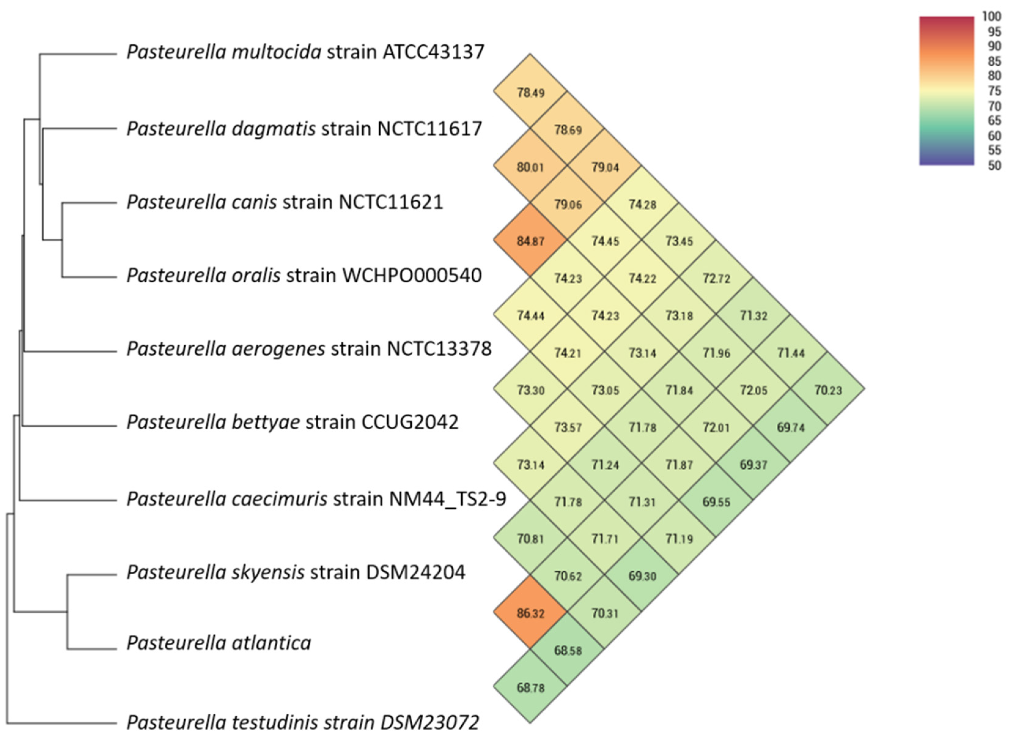
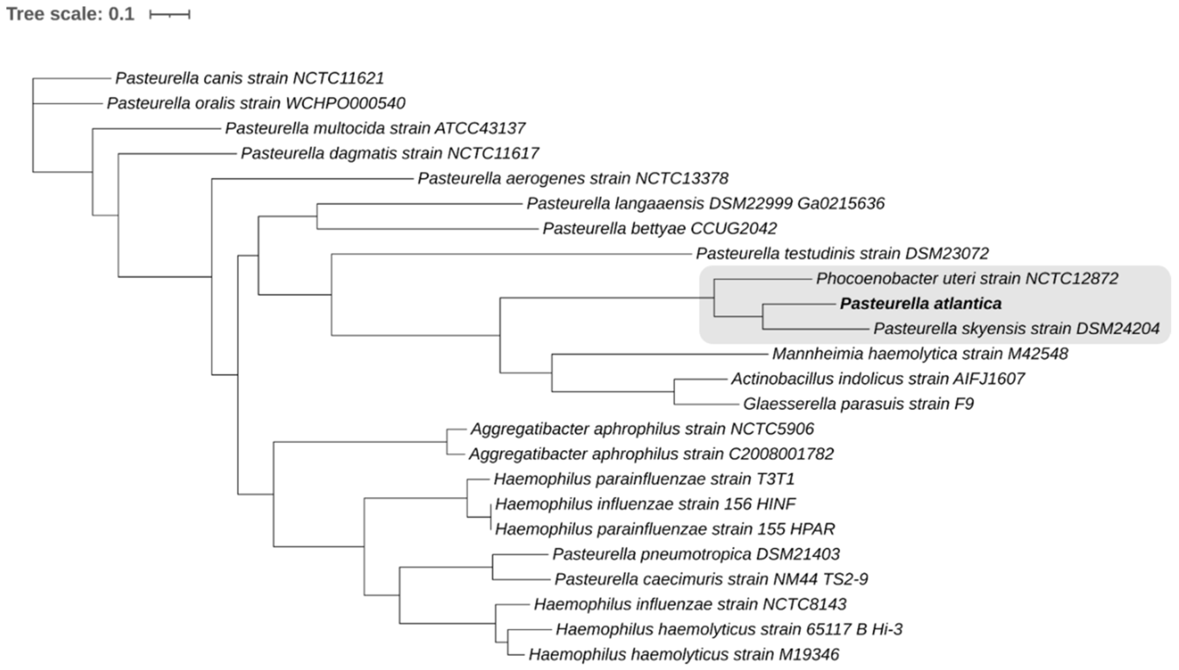
Twenty-three Pasteurellaceae genomes (Table 1) were included for phylogenetic analyses.
In order to evaluate similarity among whole genomes, Orthologous Average Nucleotide Identity Tool using OrthoANI [43] was used to provide reliable and fast assessment of average nucleotide identity (ANI) for taxonomic classification purposes. A Similar Genome Finder with high similarity parameters (max hits: 50; p-value threshold: 1; distance: 0.5) utilizing Mash, a fast genome distance estimation tool [44], mounted on the Pathosystems Resource Integration Center (PATRIC) platform (https://patricbrc.org/ (accessed on 3 June 2021)) [45,46] was also used to assess the genomic similarity of P. atlantica to the bacterial database.
PATRIC [45,46] was used to construct a whole-genome codon-based tree built on 500 single-copy genes [47] present in all genomes studied (Table 1). Both amino acid and nucleotide sequences were aligned using MUSCLE [48] and the codon align function of Biopython [49], respectively. The concatenated alignments of protein and nucleotide sequences were used in order to generate a RAxML method-based phylogenetic tree with branch support values determined by 100 rounds of rapid bootstrapping [50,51]. The tree was visualized using iTOL online platform [52].

2.3. Analysis of Genomic Regions and Reverse Vaccinology Approach

The novel genomes were screened against major virulence factor databases. Specifically, the proteome of P. atlantica was blasted against VFDB [53] and VICTORS [54] using an e-value of 1 × 10⁻¹⁰. A moderate e-value was applied to avoid false positive hits while identifying as many presumptive virulence factors as possible. The prediction was complemented by screening against PATRIC_VF [55], the integrated virulence factor database inside the PATRIC platform (https://patricbrc.org/ (accessed on 3 June 2021)) [45,46]. Subcellular localization for all presumptive virulence factors was examined by PSORTb 3.0 [56] to evaluate putative interaction with the extracellular environment (localization score threshold >7.5). Proteins with both extracellular and outer membrane subcellular localization were imported to SignalP server [57] in order to further assess any known types of signal peptides as well as the pathways in which they are involved during their secretion or anchoring process (threshold >90%). The detection of possible adhesion-related functionality was tested in SPAAN [58]. A Protein Fold Recognition Server, Phyre2 [59], was applied to obtain insights on structure and folding properties of presumptive virulence factors using confidence level of above 97%.
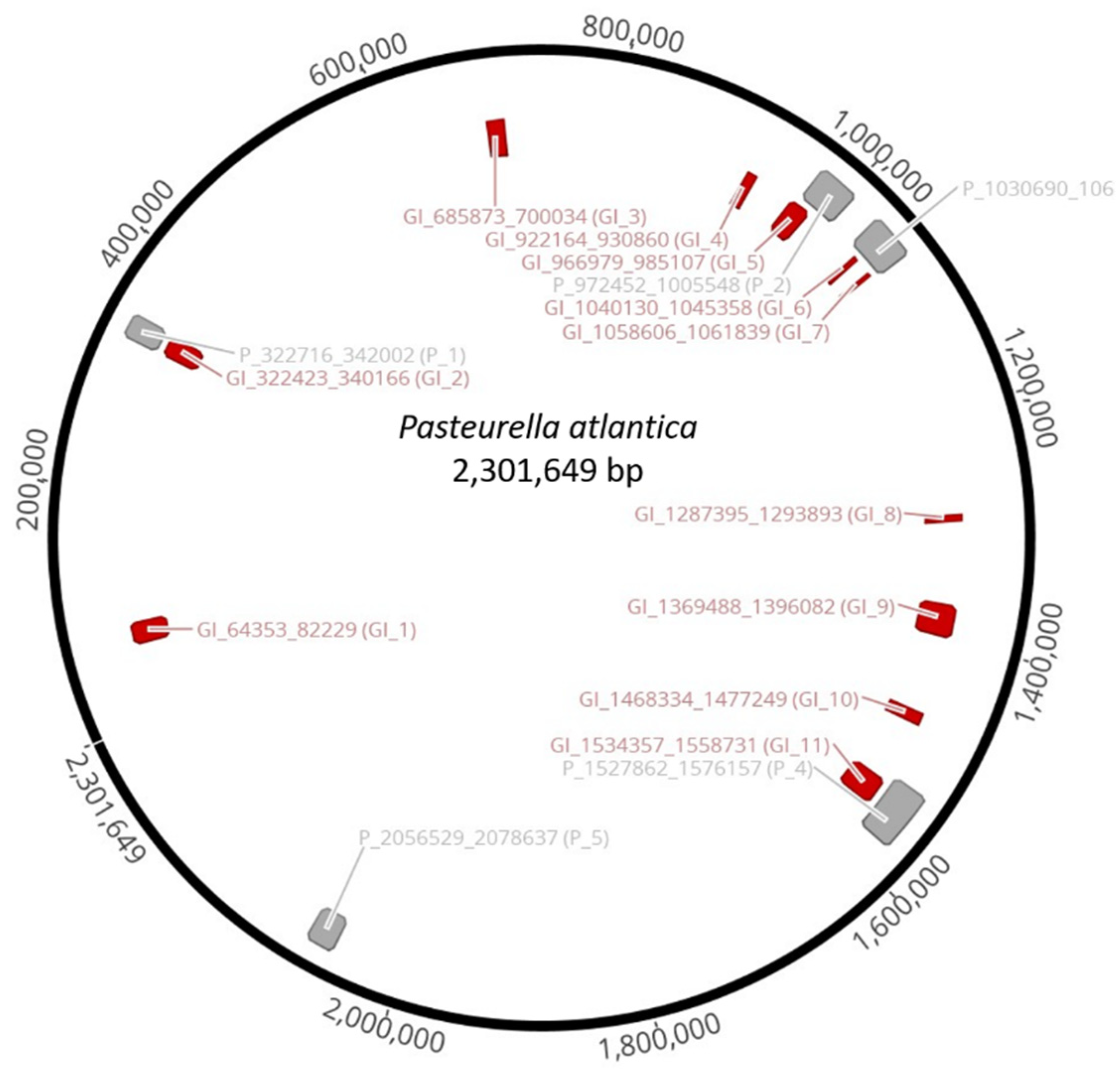
Clusters of genes of probable horizontal origin, known as genomic islands (GIs), were identified by IslandViewer 4, version 4; Simon Fraser University: Canada, 2017 [60]. Inducible prophages were detected using the prophage finder tool, PHASTER [61], while VIRFAM [62] was used to classify intact prophages.
In line with reverse vaccinology principles, the identification of the strongest virulence candidates was deduced through Dynamic Vaxign Analysis, Vaxign [63], a software platform that has been developed and dedicated to vaccine design. The pipeline of Vaxign has integrated PSORTb [56] and SPAAN [58] so it can also be used to validate the previously identified virulence factors. Vaxign-ML [20] was also included in the reverse vaccinology analysis as a machine learning model to improve the prediction of bacterial protective antigens. VaxiJen [64] complemented the analysis to corroborate that the finally selected candidates for vaccine development also can function as protective antigens, which is a prerequisite for vaccine development.

2.4. Functional Studies

2.4.1. Processing Bacteria for qPCR Analysis

Cultures of P. atlantica (UiBP1-2013) used for the in vitro exposure experiment (Section 2.4.5) were cultivated as described in Section 2.1, harvested in the late exponential growth phase (18 h post inoculation) and centrifuged at 2500× g (Beckman Coulter Allegra X-15R) for 15 min at 4 °C. For the gene expression analysis during bacterial cell proliferation in growth medium, 1 mL samples were harvested 14, 16, 18, and 20 h after inoculation. Bacterial cell counts were measured at harvest using a cell counter (CASY Model TT (Innovatis) and CASY worX version 1.26) followed by centrifugation at 4000× g (Beckman Coulter Allegra X-15R) for 10 min at 4 °C. For both methods, the growth medium was then discarded, and the samples were stored on ice.
Total RNA was immediately extracted using the Bacterial RNA Kit (E.Z.N.A., Norcross, GA, USA) according to manufacturer instructions. The RNA was then DNase-treated (Sigma-Aldrich, Saint-Louis, MO, USA), converted to cDNA using qScript cDNA Synthesis Kit (Quantabio, Beverly, MA, USA), and stored at −20 °C.

2.4.2. qPCR

Each qPCR reaction contained a volume of 25 μL and consisted of 12.5 μL 2× SYBR Green JumpStart Taq Ready Mix (Thermo-Fisher Scientific, Waltham, MA, USA), 1 μL each of the forward and reverse primers (10 μM final working concentration, Table 2), 0.5 μL of RNase and DNase free water (Sigma-Aldrich), and 10 μL of cDNA (concentration depended on the specific analysis). A C1000 Touch thermal cycler with CFX96 Real-Time System (Bio-Rad, Oslo, Norway) was used for qPCR, with the following cycle conditions: 94 °C for 5 min followed by 40 cycles of 94 °C for 15 s and 60 °C for 1 min. Melting curve analyses were performed after each run (60 to 92 °C at a rate of 1 °C/5 s) to ensure that the specificity of the primers and the qPCR products were visualized on a 2% agarose gel. Three parallel reactions were performed for each gene, and negative controls excluding cDNA (NTC) and cDNA reactions without reverse transcriptase (NRT) were included for all master mixes. The gene expression levels were calculated by the ΔΔCt method [65].

2.4.3. Primer Design and Validation

Genes considered for this work were based on the results from an analysis of genomic regions and potential vaccine targets. The genes selected as reference genes were rpoD and gyrA. The target gene selected was the putative uncharacterized protein (<Hia>) (Table 2).
qPCR assays were designed using the software Primer Premier version 6.24 (Premier Biosoft, San Francisco, CA, USA). The five highest rated assays for each target sequence were then chosen for testing. The length of the amplicons was kept between 100 and 250 bp for optimal amplification efficiency. The specificity of the primers was confirmed by qPCR (20 ng of cDNA used in each qPCR reaction), and product size was observed by electrophoresis on 2% agarose gels. All of the qPCR assays produced single amplification products. The best assay for each target gene based on Cq value, non-template controls, melting curves, and the results of electrophoresis were then chosen for further work. The resulting primers used for qPCR are listed in Table 2.

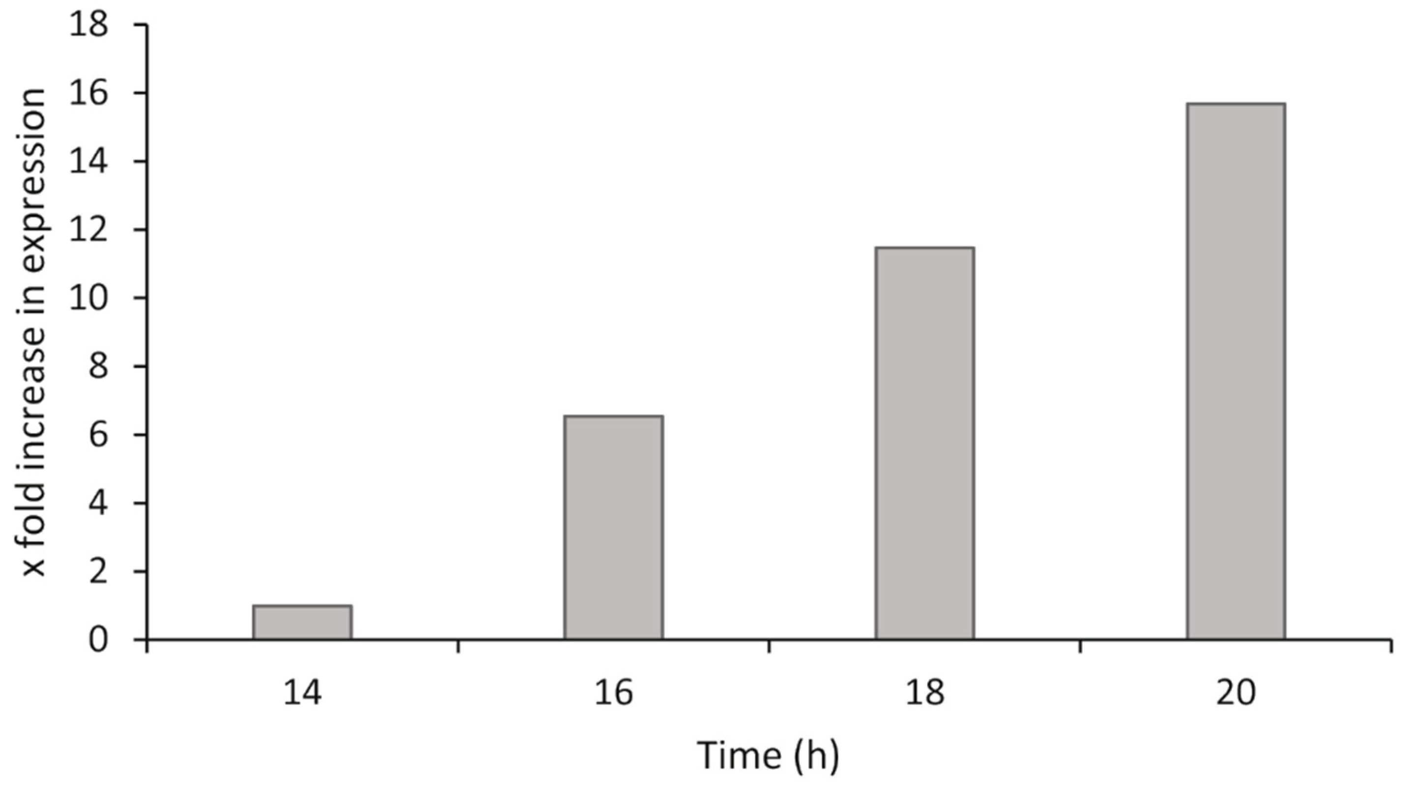
2.4.4. Bacterial Exponential Growth Phase Analysis

P. atlantica was grown as described in Section 2.1. At different time points after inoculation, the bacteria were harvested, the RNA was isolated, and cDNA synthesis was performed as described in Section 2.4.1. The synthesized cDNA was diluted across a twofold dilution series to give a range from 10 ng/µL to 0.625 ng/µL of cDNA for both the target and reference genes in qPCR (100 ng to 6.25 ng in each qPCR reaction). The relative gene expression was calculated by the ΔΔCq method using the most suitable dilution from the range tested, and comparisons of gene expression were made to the lowest stable time point of the analysis.

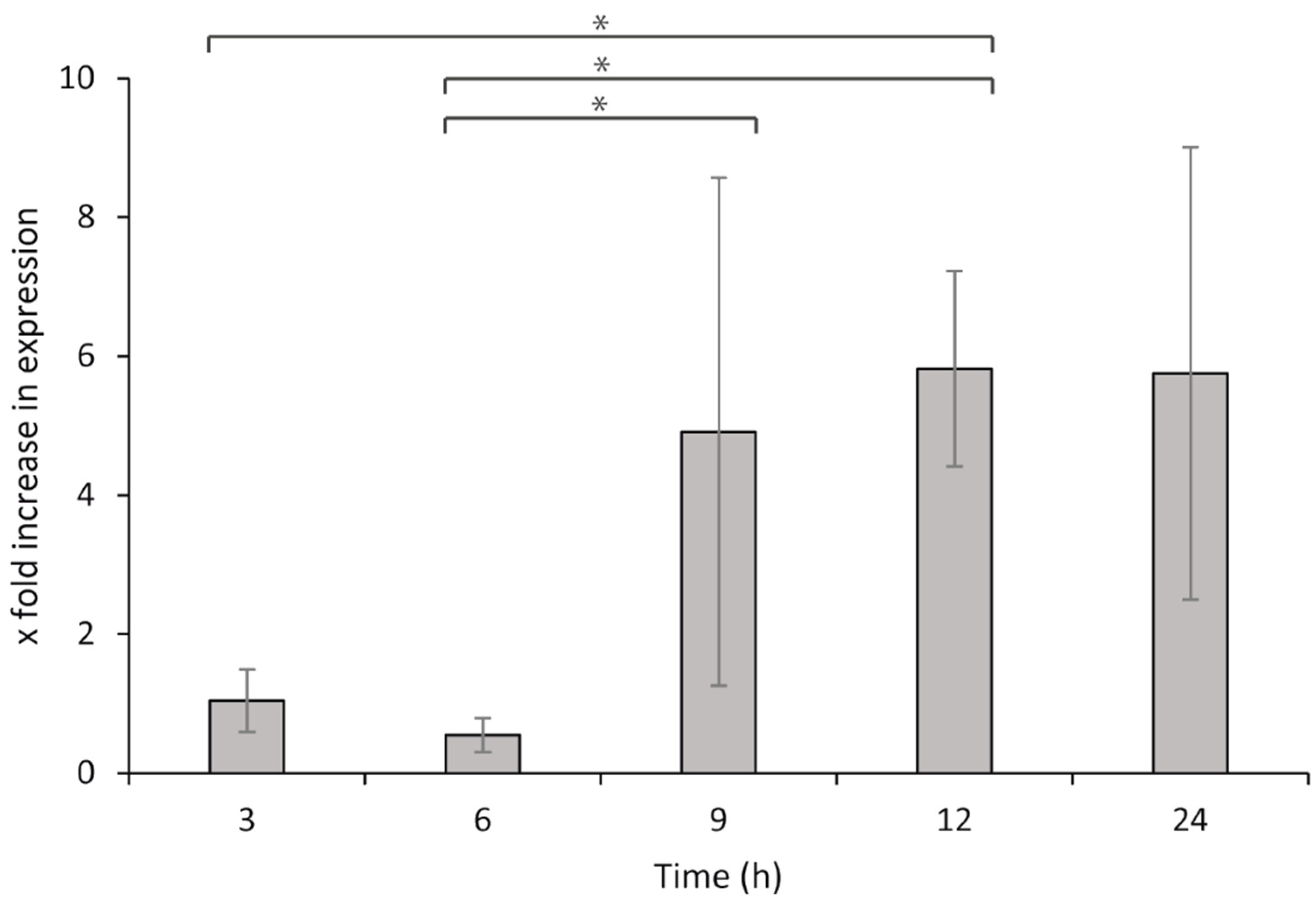
2.4.5. Head Kidney Leucocyte (HKL) Exposure Analysis

Four lumpsuckers were quickly netted and killed by a sharp blow to the head. Leucocytes were isolated from the head kidney on discontinuous Percoll gradients as described previously [66] with the following modifications. The supplemented L-15 medium did not contain gentamicin sulphate since the cells were to be exposed to viable P. atlantica. Additionally, resuspension of the isolated leucocytes was performed in L-15 supplemented with 5% fetal calf serum (L-15/FCS). The leucocytes were counted in a CASY-TT Cell Counter TM (Innovatis AG), and the viability (95%) and aggregation factor (1.2) of the cells were determined. The concentration of the isolated leucocytes was then adjusted to 3.3 × 10⁶ cells mL−1 in L-15/FCS, and 250 μL volumes were added to each well of the 24-well Nunc plates (approximately 8 × 10⁵ cells per well) (Thermo-Fisher Scientific) and incubated overnight at 15 °C prior to exposure to P. atlantica.
A late exponential phase (18 h) culture of P. atlantica was prepared as described in Section 2.4.1, re-suspended in L-15/FCS, and adjusted to 1.5 × 10⁸ bacteria mL−1. Volumes of 250 μL were then added to each well (approximately 4 × 10⁷ bacteria per well). Sterile L-15/FCS medium was supplied to the leucocytes and used for the non-challenged controls. The cells were then incubated at 15 °C, and samples were collected at 3, 6, 9, 12, and 24 h after exposure. Samples were collected from the wells and centrifuged to remove all media prior to storage at −80 °C, until the total RNA was isolated, DNase treated, and converted to cDNA as described above. For qPCR, 80 ng per reaction was used as the template for both the target and reference genes.

2.4.6. Statistical Analysis

The relative gene expression during the HKL exposure was calculated by the ΔΔCq method [65] and comparisons were made to the negative controls (bacteria without leucocytes). The results were analyzed using two-way ANOVA and Fisher’s LSD for post hoc tests, and differences were considered significant when p < 0.05. All statistical analyses were carried out using SigmaPlot version 12 (Systat Software, San Jose, CA, USA).

3. Results

3.1. Novel Sequenced Genome and Accession Numbers

The length of the PacBio sequenced P. atlantica genome (NVI 9100) is 2,301,649 bp, with a GC content of 42.1%, and 2197 coding regions (CDS) were predicted. The length of the Illumina sequenced P. atlantica genome (UiBP1-2013) is 2,260,408 bp.

3.2. Phylogenomics and Taxonomic Classification

The P. atlantica strains were isolated from Cyclopterus lumpus in a previous study where preliminary phylogenetic analysis was performed based on 16S rRNA and rpoB gene sequencing [4]. To assess pairwise similarity percentages among Pasteurella species, nine different publicly availably Pasteurella species genomes (Table 1) and P. atlantica were analyzed using the OrthoANI tool (Figure 1).
When compared to our genome of interest, P. skyensis shows the highest OrthoANI values (86.32%) whereas the other species generated much lower values (70 to 72%).
Apart from Pasteurella genus, a genomic analysis of P. atlantica against the entire publicly available bacterial database, based on whole genome sequences, according to Similar Genome Finder, returned the P. skyensis strain DSM 24204 as the only similar genome at a genomic distance of 0.096 (Mash distance threshold: 0.1). The second most similar genome at a genomic distance of 0.145 was the Ph. uteri strain NCTC12872, and that was shown only when the threshold increased up to 0.5 (Figure 1). A bigger number of Pasteurellaceae family bacteria were found to be less similar, at genomic distances of >0.2, and for the sake of further analysis, representative genomes have been included in Table 1 as well. Substantially low-similarity levels compared to the bacterial database constitute an additional piece of evidence supporting P. atlantica as a novel species, knowing that a pairwise Mash distance of at least ≤0.05, approximately corresponding to an ANI of ≥95%, is taken for granted among conspecific bacterial genomes [44].
Multiple codons through the entire genomes of Table 1 were used as genetic markers to finalize the taxonomy of P. atlantica (Figure 2).
According to the genome-wide analysis, P. atlantica clusters together with P. skyensis and Ph. uteri, forming a robust phylogenetic group supported by 100% confidence level (highlighted in Figure 2). Both monophyletic taxa are phylogenetically distinct since their nodes bear 100% support values.

3.3. Genomic Elements

The sequence of P. atlantica revealed mobile elements including genomic islands and prophages that should further be analyzed due to their possible role in pathogenicity [67,68,69].

3.3.1. Genomic Islands and Prophages

Bioinformatic analysis on the genome of P. atlantica illustrated eleven genomic islands (GIs) (Table 3) with various properties.
GC content of the GIs span between 29.4 and 36.6%. They encompass 207 coding sequences (CDS), approximately 10% of the P. atlantica total proteome, and many of them were attributed to virulence-related or broadly proteolytic activity functions. The most prominent putative virulence functions belong to CDS such as RTX-like toxin (GI_1), putative hemolysin toxin of HlyC family (GI_1), heme-binding protein (GI_4), bacterial peptidoglycan degrading enzyme of NIpC/P60 (GI_6), spermidine-putrescine binding (GI_8), acetyltransferase of gnat family (GI_9), ctx rstr-like repressor (GI_9), two-partner secretion (TPS) system protein (GI_10), and toxin-antitoxin system proteins (GI_11).
The genome of P. atlantica (NVI 9100) harbors five different prophages that were in silico predicted to be intact, hence inducible (Table 4).
P_1 and P_3 were found to be somewhat related to Enterobacteria phages, whereas P_2, P_4, and P_5 resemble prophages that have already been described in other Pasteurellaceae family bacteria [70,71]. According to BLAST sequence similarity, it was only possible to match 4 out of 26 CDS (7.7%) present in P_1 with any known phage genome implying that this is a quite novel prophage, uniquely present in the genome of P. atlantica. Similarly, P_3 seems to be a unique prophage as only 9 of its 49 CDS (18.4%) aligned with Escherichia phage D108. On the other hand, P_2 carried 9 of 40 CDS (22.5%) of Mannheimia phage vB_MhS_535AP2 while P_4 has homologs of 24 out of 57 CDS (42.1%) that were present in Mannheimia phage vB_MhM_587AP1. Last, P_5 has 23 out of 28 CDS (82.1%), resembling Hemophilus phage SuMu more, which was also anticipated due to the close phylogenetic relationships between P. atlantica and G. parasuis [40].
Interestingly, some of the predicted genomic islands and prophages do overlap in the genome (Figure 3). Specifically, the entire GI_2 is part of P_1, part of GI_5 is included in P_2, both GI_6 and GI_7 belong to P_3, while GI_11 is a part of P_5. Hence, there is a prominent connection between prophages and genomic islands that may be directly linked to the virulence properties of the bacterial host, with the former inciting the mobilization of the latter.

3.3.2. Virulence Factors

The proteome of P. atlantica consists of 2197 protein coding sequences (Supplementary Material S1), which were screened against an integrated matrix consisting of the VFDB, VICTORS, and PATRIC_VF databases to generate 565 unique amino acid sequence hits after discarding overlapping hits (Supplementary Material S1). This corresponds to approximately 26% of the 2197 predicted coding sequences. However, significant uncertainty needs to be removed from the analysis to achieve a better focus on the essential virulence factors present in the species. Subcellular localization of presumptive virulence factors showed that approximately 12% (70 out of 565) are presumably either extracellularly secreted (21) or with outer membrane surface exposure (49), setting these proteins as primary candidates for the pathogenicity of P. atlantica. The amino acid sequences of the predicted proteins accompanied by their most similar protein sequence GI number (SCL-BLAST), subcellular localization, adhesion, and transmembrane (TM) helices probabilities are reported in Supplementary Material S1. According to their amino acid sequences, 17 out of the 70 virulence factors with extracellular and outer membrane subcellular localization were the most critical for mediating bacterial invasion, such as adhesins with probabilities higher than 75%. Adhesins correspond to 0.8% of the total proteome (Figure 4), and since their expression may be a key determinant of the success of the infection process [72], they could play an imperative role in the pathogenicity of P. atlantica (Table 5).
The strongest virulence factor candidate is a novel uncharacterized protein, the function of which would have passed unnoticed without examining its protein structure. Therefore, PDB predictions based on protein fold recognition were included. All PDB predictions reported in Table 5 were above the 97% confidence level and encompassed major virulence components such as hemolysins, toxins, and pili subunits. Specific secretory signal peptides were detected in only 2 out of 17 adhesins, intended to be transported by Sec translocon followed by Signal Peptidase I cleavage (Sec/SPI) to be released to the host.

3.3.3. Vaccine Targets

All 70 CDS with extracellular and outer membrane localization were assessed as potential subunit vaccine candidates. However, apart from adhesion probability, additional factors such as transmembrane helices and orthologue analysis need to be considered for calculating their Vaxign-ML score, which was the metric applied to define their suitability as targets for vaccine development. All vaccine targets with Vaxign-ML scores higher than 99.5% are presented in Table 6, and they are therefore suggested as the most promising vaccine targets. All of the exact scores as well as further details about the remaining 60 extracellular and outer membrane CDS are included in Supplementary Material S1.
Since adhesion probability is not the only criterion for vaccine development, 6 out of the 10 CDS reported in Table 5 are potential virulence factors (Table 6). The list can further be confined by using VaxiJen as a tool to predict the protective antigenic properties of the vaccine targets. Only the putative uncharacterized protein and the Hep/Hag repeat protein had a score >80%, claiming that their antigenicity levels are high enough to trigger the host’s immune response. However, the putative uncharacterized protein CDS not only was ranked first in both tables, scoring the highest Vaxign-ML (100%) and adhesin probability (92.5%), respectively, but also was characterized as a protective antigen, findings that render it the most significant candidate for virulence and potential vaccine development.
According to its amino acid sequence, BLASTP identified it as a YadA-like family protein, a collagen-binding adhesin originally characterized in Yersinia enterocolitica [73], that is also present in the genome of P. skyensis. However, although the query coverage was 100%, the identity rate was only 58.23%. Based on the predicted structure of the protein, Phyre2 [59] identified it as a membrane protein for cell adhesion similar to Hia (Table 5 and Table 6), which is also a major adhesin expressed by H. influenzae [74]. Due to the prominent role that this uncharacterized membrane/cell adhesion protein seems to play in the virulence of P. atlantica, it was selected for further evaluation of its expression levels against lumpsucker head kidney leucocytes.

3.3.4. Functional Analysis of the Major Virulence Factor

Expression of <hia> in Bacterial Exponential Growth Phase

The predetermined time points for measurement were chosen based on growth curves of P. atlantica to coincide with the exponential growth phase of the bacteria. Furthermore, from the range of cDNA dilutions tested, 50 ng of cDNA per qPCR reaction yielded the clearest and most consistent results, and this dilution was used to calculate the <hia> expression. Out of the two reference genes tested, gyrA gave the most stable results compared to rpoD (Table 7) and was therefore used to calculate the relative expression of <hia> during cell growth and leucocyte exposure studies. This highlights the importance of testing various reference genes for evaluation and stability for expression studies. In this study, although rpoD seemed to be a stable gene to use for analysis when primer assays were evaluated, experimental situations proved otherwise.
The expression level of <hia> changed over time, when analyzing samples collected at different time points during the exponential growth phase. The expression levels were upregulated by 6.5×, 11.5×, and 15.7× at 16, 18, and 20 h, respectively, compared to the expression level at 14 h (Figure 5). The expression levels also mirror the exponential increase seen in bacterial numbers from previous work performed on growth curves for P. atlantica [28].

Head Kidney Leucocyte (HKL) Exposure Analysis

The expression levels of <hia> increased with time, with the highest levels recorded at 9 and 12 h after leucocyte exposure (Figure 6), with significantly elevated expression levels compared with those in the early stages of the exposure (3 and 6 h after exposure). At 24 h after exposure compared to 3 and 6 h after exposure, the bacteria would have entered the lag phase of growth, and the expressions of both the reference and target genes are therefore not comparable with those from earlier time points. Furthermore, statistical analysis also showed that HKL from individual fish did not have an impact on the expression of <hia> (p = 0.33).

4. Discussion

The isolates of P. atlantica used in this work were isolated and reported as the causative agent of confirmed outbreaks of pasteurellosis in farmed lumpsuckers from Norway.

4.1. Phylogeny Reconstruction

Alarcón et al. [4] carried out preliminary phylogenetic analyses that included the isolates used in this study, using the 16S rRNA and rpoB genes, and it was suggested yet not concluded that the isolates might represent a novel species or subspecies. After examining the existing phylogeny of the Pasteurellaceae family, which is based on 16S rRNA genes, it can be deduced that using this molecular marker does not sufficiently resolve the true phylogenetic relationships, since it reflects rather incongruent phylogenetic relationships among its members. The available 16S rRNA based phylogenetic tree of the Pasteurellaceae family provides clear evidence that the polyphyletic nature prevails in the taxonomy of the family [75] and it has already been proven that there is room for improvement on molecular markers [76]. Despite ongoing research, the current phylogeny of the Pasteurellaceae family cannot be considered resolved according to the evolutionary principle of monophyletic taxa formation. Phylogenomic and molecular demarcation studies have been performed both on the major genera of the family such as Pasteurella, Hemophilus, and Actinobacillus [77] and on the reclassification of individual species such as the case of Hemophilus parasuis to G. parasuis [40].
Hence, the classification of P. atlantica in the current study was based on a dataset that includes representative species from both Pasteurella and other genera that belong to the Pasteurellaceae family to unravel a comprehensive taxonomy for P. atlantica. P. multocida is the most thoroughly studied member of Pasteurellaceae due to its high pathogenicity in a broad range of livestock species [78,79]. The observed orthologous average nucleotide of 86.32% corroborates that P. atlantica and P. skyensis represent two different species. Whole genome-based phylogeny validated the novelty of the studied genome and its accurate taxonomic placement. Since P. skyensis and Ph. uteri are the only strains of marine origin out of the entire bacterial database, the habitat seems to be a strong differentiating factor for this taxon of Pasteurellaceae. Second, the host species of the bacteria in the marine cluster likely play a role for the speciation process. P. atlantica and P. skyensis are the closest relatives and both infect marine teleosts [35], whereas Ph. uteri isolated from a marine mammal is classified slightly more distant [36]. The major setback in this case is the lack of data in sequenced aquatic Pasteurellaceae, a fact that makes it quite precarious to formulate any solid conclusions regarding the trait of virulence. However, the highly emerging incidence and pathogenicity of Pasteurella in aquaculture has already brought [4,80] and will definitely bring more genomic data to the scientific community.

4.2. Virulence Genes

The scarcity of aquatic Pasteurella genomes, particularly from the aquaculture environment, highlights the significance of these P. atlantica genomes in exploring genomic elements, including putative virulence factors, antimicrobial resistance genes, and their potential implications for devising precaution strategies such as vaccine development in aquaculture. The abundance of mobile elements such as GIs (11) and prophages (5) in P. atlantica further enforces its high pathogenicity that has already been documented in previous studies [4,6,28]. GIs and prophages are very often found in pathogenic bacteria and have consistently been reported as major contributors of virulence factors to their bacterial hosts. Five out of the seventeen strongest candidate virulence factors of P. atlantica are in GIs and/or prophages, which in total account for roughly 10% of the genome. This finding is in line with the current literature, which supports the potential of bacterial mobilomes to harbor significant pathogenicity traits. A recent comparative genomic analysis of Vibrio anguillarum, a highly virulent aquaculture pathogen, revealed that 64 GIs that belonged to the accessory genome of the species were present in six out of nine of the most virulent strains [67]. In the same study, the role of prophages was also quite prominent, while it was recently proven in another study that marine Vibrio harbors a large number of prophage-encoded virulence factors [81]. In the pangenome analysis of P. multocida, GIs were also characterized as one of the most important mobile components not only as sources of virulence factors but also as facilitators of foreign genes acquisition. Prophages were again highlighted as core elements of the mobilome, and it was speculated that they may serve as precursors for the formation of GIs [69]. Additionally, facultative intracellular pathogens, such as P. atlantica [6,28,82], usually demonstrate quite variable genomes with a fourfold larger genomic content of mobile DNA such as transposable elements and prophages, when compared to obligate intracellular bacteria [83].
Mobile elements are characterized by a high frequency of transfer in the environment through the mechanism of horizontal gene transfer (HGT). HGT can be phage-mediated and can facilitate the dissemination of virulence-associated GIs within and between microbial communities [84], and the presence of a pathogenicity island can potentially define virulence in bacterial pathogens [85]. Apart from sophisticated algorithms [60,61], simpler methods that are based on the distinct GC content of horizontally transferred genes compared to that of the bacterial chromosome have been suggested for the prediction of GIs and prophages [86]. For instance, the pathogenicity islands in Salmonella typhimurium (SPIs) have lower GC content than the rest of the bacterial genome [87]. According to the in silico analysis of our study, the average GC content for GIs and prophages were 34.5% and 35.6%, respectively, in both cases lower than 42.1%, which is the average GC content of the P. atlantica genome.
Recent progress in in silico analysis tools allowed us to recognize and assess potential virulence factors by exploring the novel genome of P. atlantica. Such methodologies constitute fundamental principles of RV, which facilitate vaccine development, while turning extensive, time consuming, and generalized lab work into target-oriented functional analysis and in vitro/in vivo trials. It was not until 2006 when the first standalone RV program designated as NERVE [88] was published, whereas great progress as well as comparative analysis have been achieved and performed in the RV field ever since [89]. The cornerstone of virulence factor prediction has been the determination of subcellular localization of bacterial proteins. Proteins that are extracellularly secreted or exposed on the bacterial surface have good chances to play a role in bacterial virulence.

4.3. Adhesins

Adhesins, for instance, which can facilitate the colonization of pathogens to mucosal or other biotic surfaces and which are essential for bacterial pathogenesis and survival may be prominent virulence factors and suitable candidates for vaccine development [72,90]. Screening of the P. atlantica genome for gene targets that can be further functionally evaluated as virulence factors generated the 17 strongest candidate genes that may have implications for bacterial virulence, based on subcellular localization and predicted adhesion properties.
Previously published bath, cohabitation, and intramuscular and intraperitoneal challenge trials that included the current P. atlantica isolate (UiBP1-2013) illustrated high and acute mortality rates, potentially indicating that extracellular proteins may have a substantial role in the infection dynamics [28], an interim conclusion that is supported by present findings. In our analysis, a putative uncharacterized protein ranked first as both a putative virulence factor and a vaccine candidate.
According to the in silico analysis, the role of this protein was directly linked to major adhesins YadA and Hia in Yersinia and H. influenzae, respectively, both of which are essential for successful infection of their hosts via human epithelia tissues [73,74,91]. Although their amino acid sequences were not identical, there was a clear connection based on query coverage and confidence level, which in both cases were 100%.
Trimeric autotransporters (TAAs) are a family of non-fimbrial, homotrimeric bacterial adhesins that are secreted by the Type 5c secretion system (T5cSS) and for which the main function is the attachment of pathogenic Gram-negative bacteria to hosts and abiotic surfaces. Yersinia adhesin A (YadA) is the most extensively studied member of this family, while others include H. influenzae adhesin (Hia) and Neisseria adhesin A (NadA).
The type 5c secretion system is of great importance in the development of multivalent recombinant and subunit vaccines, as it enables proteins to be exposed on the bacterial outer membrane, making them potential vaccine targets [92]. This could then also be used as the basis for further work to elucidate the structure of the Hia-like protein investigated in the current study.
YadA showcases the invasive nature of Y. enterolitica and Y. pseudotuberculosis, which colonize the intestine by adhering to the intestinal mucosa [93]. Immunologically, YadA has been shown to be a highly immunogenic antigen. In fact, Tahir and Skurnik (2001) [93] found that immunizing mice with live bacteria raised antibodies against only epitopes located in the N-termini, whereas using inactivated bacteria raised antibodies against other epitopes as well. In addition, a study [94] found that mice immunized using a recombinant YadA vaccine had higher survival compared to those vaccinated with inactivated Y. pseudotuberculosis.
Hia is an adhesin found in 25% of clinical non-typable strains of H. influenzae (NTHi). It mediates adherence to host cells and causes various diseases in humans, including respiratory tract infections and meningitis [95]. Hia differs slightly from other TAAs and is thought to represent a new sub-family. It too is highly immunogenic, however, and antibodies were strongly induced following natural infection observed in humans. Furthermore, opsonophagocytic antibodies were generated in serum in guinea pigs and mice [96]. However, due to its prevalence in so few strains and despite its high antigenicity, it cannot be considered a vaccine target on its own but in combination with High Molecular Weight adhesin 1 (HMW1), High Molecular Weight adhesin 2 (HMW2), and outer membrane vesicles (OMVs). In this way, a vaccine containing the three adhesins covers 95% of NTHi and confers better protection [97].
The significantly higher expression levels of <hia> detected in the current study following in vitro exposure to lumpsucker leucocytes after 9 and 12 h together with the in silico results strengthen its candidacy as both a major virulence factor and a potential target for subunit protein antigen, mRNA, or DNA vaccine development.
The expression of <hia> in the absence of a host correlates with previously observed increases in bacterial growth rates [28] and indicates that the adhesin is typically expressed during bacterial growth. In the exposure experiment, bacteria were harvested from the growth medium at 18 h after inoculation when both bacterial growth and <hia> expression were at peak levels and combined with HKL in cell culture medium, which does not favor bacterial growth. The results from this experiment then demonstrate that the expression of <hia> increases significantly in the presence of the host, following an initial period of acclimation (up to 6 h, as seen in Figure 6). This implies that the presence of host leucocytes serves as a trigger for the bacteria to significantly increase the expression of adhesins (between 6 and 9 h after exposure) and shows that the increased expression is due to the presence of the host and not an increase in bacterial cell numbers. Furthermore, it indicates bacterial cell attachment to leucocytes as an initial step towards infection, as previously suggested by Ellul et al. (2019) [6].

4.4. Vaccine Development

Vaccines targeting adhesins can confer protection by two major mechanisms, one of which is by inducing neutralizing antibodies, which attach to adhesins and prevent attachment and subsequent infection. The other involves inducing opsonizing antibodies, which attach to adhesins and increase recognition of the pathogen by Fc receptors on phagocytes, leading to phagocytosis, or by activation of the complement system [98], as shown by Winter and Barenkamp [96] in their experiments with Hia.
Ellul et al. [6] recently assessed the efficacy of formalin-inactivated P. atlantica vaccine in lumpsucker, and although immune response and antibody production were prominent, the protection levels provided by the vaccine were relatively weak. Inactivated bacterin is the most common and traditional type of vaccine used in aquaculture; however, alternatives such as subunit recombinant, mRNA, or DNA vaccines may be the next step in solving the problem [99]. This is especially the case with the recent advances in human mRNA vaccines against SARS-CoV-2, which point to potential avenues for fish vaccines. These vaccines can be developed using various techniques, including harvesting and purifying the target directly from the pathogen, or using recombinant expression vectors, such as E. coli [99]. Pending further work, the former method could be a possible step forward in the case of P. atlantica as high expression rates were observed in bacteria in their exponential growth stage.
Some of the most successful recombinant protein subunit vaccines in the aquaculture industry include those against IPN (MSD Animal Health) and ISA in Chile (Centrovet). DNA vaccines have also been produced for use in Atlantic salmon against IHN in Canada and against SPD in Norway (Elanco). Subunit vaccines against the bacterial fish disease furunculosis in Oncorhynchus mykiss [100] have been tested. All targets for the vaccine development were identified in silico, employing the RV principles and found to be protective. Recombinant protein production has already been used in order to protect farmed fish against infections by V. anguillarum, Edwardsiella tarda, Aeromonas hydrophila, and Francisella orientalis [101] in all cases leading to the suggestion of potential vaccine candidates [102,103,104]. Following these lines, our study highlights the potential that in silico analysis may have in the future formulation of a novel vaccine against pasteurellosis.
Commercial vaccines are available only for a few infectious diseases in aquaculture, and given that the needs for fish and shellfish protein as well as the number of cultured aquatic species are constantly increasing, novel vaccine development will be essential to guarantee improved animal welfare and a safe, sustainable, and healthy product.

5. Conclusions

In this study, we report the first fully sequenced genomes of P. atlantica, an emerging pathogen in Cyclopterus lumpus in Norwegian and Scottish aquaculture. The wide phylogenetic distance from the closest bacterial relative, P. skyensis, and the very few available Pasteurellaceae members of aquatic origin highlight the need for intensification of sequencing efforts that should be focused on Pasteurella strains from the aquaculture environment. In silico genomic analysis revealed numerous target genes that may be responsible for the virulence of this species, and a major role was attributed to mobile elements such as prophages and GIs. Functional analyses showed that the expression levels of an uncharacterized protein increased significantly when P. atlantica was exposed to leucocytes from lumpsuckers, complementing the in silico analysis by designating a prospective virulence gene and by promising candidate for vaccine development. Further assessment of virulence in larger scale as well as formulation of experimental vaccines will shed more light on devising a precautionary strategy against a serious emerging disease in aquaculture.

Supplementary Materials

The following are available online at https://www.mdpi.com/article/10.3390/microorganisms9061215/s1, Supplementary Material S1: Database with an extensive description of the sequences from the proteome-filtering process (virulence factors and adhesins)

Author Contributions

Conceptualization, A.R. and H.I.W.; methodology, R.M.E., C.F., P.G.K., and A.R.; software, R.M.E., C.F., S.G., G.T.H., and P.G.K.; validation, R.M.E., C.F., P.G.K., and A.R.; formal analysis, R.M.E., C.F., and P.G.K.; investigation, R.M.E. and P.G.K.; resources, H.I.W.; data curation, R.M.E., C.F., and P.G.K.; writing—original draft preparation, R.M.E. and P.G.K.; writing—review and editing, D.J.C., R.M.E., C.F., S.G., G.T.H., P.G.K., M.M., A.R., and H.I.W.; visualization, R.M.E. and P.G.K.; supervision, P.G.K., A.R., and H.I.W.; project administration, A.R. and H.I.W.; funding acquisition, H.I.W. All authors have read and agreed to the published version of the manuscript.

Funding

This research was funded by the University of Bergen and supported by ACD Pharmaceuticals and the Norwegian Veterinary Institute.

Institutional Review Board Statement

This work was conducted according to the regulations of the Norwegian Food Safety Authority, National Assignment department, based on the EU directive (2010/63/EU) and defined as a non-regulated procedure.

Data Availability Statement

This whole genome project has been deposited at GenBank under the Accession Number PRJNA721926 for isolate UiBP1-2013 and accession number CP074346 for isolate NVI 9100.

Acknowledgments

R.M.E. thanks Harald Sæbø Lunde for his technical support during the design of the primers for the functional analyses in this work. PacBio sequencing was performed at the Norwegian Sequencing Centre (NSC) at the Department of Medical Genetics (AMG), Oslo University Hospital.

Conflicts of Interest

The authors declare no conflict of interest. The funders had no role in the design of the study; in the collection, analyses, or interpretation of data; in the writing of the manuscript; or in the decision to publish the results.

References

  1. Johansen, R. Fiskehelserapporten 2012; Veterinærinstituttet: Oslo, Norway, 2013. [Google Scholar]
  2. Erkinharju, T.; Bornø, G.; Grønbech, S.; Gulla, S.; Hansen, H. Helsesituasjonen hos rensefisk. In Fiskehelserapporten 2019; Sommerset, I., Walde, C., Bang Jensen, B., Bornø, G., Haukaas, A., Brun, E., Eds.; Veterinærinstituttet: Oslo, Norway, 2020; pp. 134–143. [Google Scholar]
  3. Nilsen, H.; Colquhoun, D.; Gulla, S. Pasteurella infeksjon hos laks. In Fiskehelserapporten 2020; Sommerset, I., Bang Jensen, B., Bornø, G., Haukaas, A., Brun, E., Eds.; Veterinærinstituttet: Oslo, Norway, 2021; pp. 98–99. [Google Scholar]
  4. Alarcón, M.; Gulla, S.; Røsæg, M.V.; Rønneseth, A.; Wergeland, H.; Poppe, T.T.; Nilsen, H.; Colquhoun, D.J. Pasteurellosis in lumpsucker Cyclopterus lumpus, farmed in Norway. J. Fish Dis. 2016, 39, 489–495. [Google Scholar] [CrossRef]
  5. Kui, S. Stamfisk screening. In Proceedings of the Rensefiskkonferansen; PatoGen Analyse: Trondheim, Norway, 2017. [Google Scholar]
  6. Ellul, R.M.; Bulla, J.; Brudal, E.; Colquhoun, D.; Wergeland, H.; Rønneseth, A. Protection and antibody reactivity in lumpsucker (Cyclopterus lumpus L.) following vaccination against Pasteurella sp. Fish Shellfish Immunol. 2019, 95, 650–658. [Google Scholar] [CrossRef]
  7. Bidmos, F.A.; Siris, S.; Gladstone, C.A.; Langford, P.R. Bacterial vaccine antigen discovery in the reverse vaccinology 2.0 Era: Progress and challenges. Front. Immunol. 2018, 9, 2315. [Google Scholar] [CrossRef] [Green Version]
  8. Ong, E.; Cooke, M.F.; Huffman, A.; Xiang, Z.; Wong, M.U.; Wang, H.; Seetharaman, M.; Valdez, N.; He, Y. Vaxign2: The second generation of the first Web-based vaccine design program using reverse vaccinology and machine learning. Nucleic Acids Res. 2021, gkab279. [Google Scholar] [CrossRef] [PubMed]
  9. He, Y.; Xiang, Z.; Mobley, H.L.T. Vaxign: The first web-based vaccine design program for reverse vaccinology and applications for vaccine development. J. Biomed. Biotechnol. 2010, 2010, 297505. [Google Scholar] [CrossRef] [PubMed]
  10. Sharma, A.; Sanduja, P.; Anand, A.; Mahajan, P.; Guzman, C.A.; Yadav, P.; Awasthi, A.; Hanski, E.; Dua, M.; Johri, A.K. Advanced strategies for development of vaccines against human bacterial pathogens. World J. Microbiol. Biotechnol. 2021, 37, 67. [Google Scholar] [CrossRef] [PubMed]
  11. Pizza, M.; Scarlato, V.; Masignani, V.; Giuliani, M.M.; Aricò, B.; Comanducci, M.; Jennings, G.T.; Baldi, L.; Bartolini, E.; Capecchi, B.; et al. Identification of vaccine candidates against serogroup B meningococcus by whole-genome sequencing. Science. 2000, 287, 1816–1820. [Google Scholar] [CrossRef] [PubMed]
  12. Masignani, V.; Pizza, M.; Moxon, E.R. The development of a vaccine against Meningococcus B using reverse vaccinology. Front. Immunol. 2019, 10, 751. [Google Scholar] [CrossRef] [PubMed]
  13. Betts, J.C. Transcriptomics and Proteomics: Tools for the Identification of Novel Drug Targets and Vaccine Candidates for Tuberculosis. IUBMB Life 2002, 53, 239–242. [Google Scholar] [CrossRef]
  14. Montigiani, S.; Falugi, F.; Scarselli, M.; Finco, O.; Petracca, R.; Galli, G.; Mariani, M.; Manetti, R.; Agnusdei, M.; Cevenini, R.; et al. Genomic approach for analysis of surface proteins in Chlamydia pneumoniae. Infect. Immun. 2002, 70, 368–379. [Google Scholar] [CrossRef] [Green Version]
  15. Wizemann, T.M.; Heinrichs, J.H.; Adamou, J.E.; Erwin, A.L.; Kunsch, C.; Choi, G.H.; Barash, S.C.; Rosen, C.A.; Masure, H.R.; Tuomanen, E.; et al. Use of a whole genome approach to identify vaccine molecules affording protection against Streptococcus pneumoniae infection. Infect. Immun. 2001, 69, 1593–1598. [Google Scholar] [CrossRef] [Green Version]
  16. Chakravarti, D.N.; Fiske, M.J.; Fletcher, L.D.; Zagursky, R.J. Application of genomics and proteomics for identification of bacterial gene products as potential vaccine candidates. Vaccine 2000, 19, 601–612. [Google Scholar] [CrossRef]
  17. Ross, B.C.; Czajkowski, L.; Hocking, D.; Margetts, M.; Webb, E.; Rothel, L.; Patterson, M.; Agius, C.; Camuglia, S.; Reynolds, E.; et al. Identification of vaccine candidate antigens from a genomic analysis of Porphyromonas gingivalis. Vaccine 2001, 19, 4135–4142. [Google Scholar] [CrossRef]
  18. Ariel, N.; Zvi, A.; Grosfeld, H.; Gat, O.; Inbar, Y.; Velan, B.; Cohen, S.; Shafferman, A. Search for potential vaccine candidate open reading frames in the Bacillus anthracis virulence plasmid pXO1: In silico and in vitro screening. Infect. Immun. 2002, 70, 6817–6827. [Google Scholar] [CrossRef] [Green Version]
  19. Kempsell, K.E.; Kidd, S.P.; Lewandowski, K.; Elmore, M.J.; Charlton, S.; Yeates, A.; Cuthbertson, H.; Hallis, B.; Altmann, D.M.; Rogers, M.; et al. Whole genome protein microarrays for serum profiling of immunodominant antigens of Bacillus anthracis. Front. Microbiol. 2015, 6, 747. [Google Scholar] [CrossRef] [PubMed] [Green Version]
  20. Ong, E.; Wang, H.; Wong, M.U.; Seetharaman, M.; Valdez, N.; He, Y. Vaxign-ML: Supervised machine learning reverse vaccinology model for improved prediction of bacterial protective antigens. Bioinformatics 2020, 36, 3185–3191. [Google Scholar] [CrossRef] [PubMed]
  21. Abraham, S.; Sharon, N.; Ofek, I.; Schwartzman, J. Adhesion and colonisation. In Molecular Medical Microbiology; Sails, A., Poxton, I., Liu, D., Schwartzman, J., Eds.; Elsevier Science: Brussels, Belgium, 2014; pp. 409–421. [Google Scholar]
  22. Moon, H.W.; Bunn, T.O. Vaccines for preventing enterotoxigenic Escherichia coli infections in farm animals. Vaccine 1993, 11, 213–220. [Google Scholar] [CrossRef]
  23. Langermann, S.; Möllby, R.; Burlein, J.E.; Palaszynski, S.R.; Auguste, C.G.; DeFusco, A.; Strouse, R.; Schenerman, M.A.; Hultgren, S.J.; Pinkner, J.S.; et al. Vaccination with FimH Adhesin Protects Cynomolgus Monkeys from Colonization and Infection by Uropathogenic Escherichia coli. J. Infect. Dis. 2000, 181, 774–778. [Google Scholar] [CrossRef] [PubMed] [Green Version]
  24. Fang, H.M.; Ge, R.; Sin, Y.M. Cloning, characterisation and expression of Aeromonas hydrophila major adhesin. Fish Shellfish Immunol. 2004, 16, 645–658. [Google Scholar] [CrossRef]
  25. Maiti, B.; Shetty, M.; Shekar, M.; Karunasagar, I.; Karunasagar, I. Evaluation of two outer membrane proteins, Aha1 and OmpW of Aeromonas hydrophila as vaccine candidate for common carp. Vet. Immunol. Immunopathol. 2012, 149, 298–301. [Google Scholar] [CrossRef]
  26. Zhu, Z.; Dong, C.; Weng, S.; He, J. Identification of outer membrane protein TolC as the major adhesin and potential vaccine candidate for Vibrio harveyi in hybrid grouper, Epinephelus fuscoguttatus (♀) × E. lanceolatus (♂). Fish Shellfish Immunol. 2019, 86, 143–151. [Google Scholar] [CrossRef] [PubMed]
  27. Jin, C.-D.; Li, M.-F. Edwardsiella tarda outer membrane protein 1 is a virulence factor and a protective immunogen. Aquaculture 2021, 539, 736596. [Google Scholar] [CrossRef]
  28. Ellul, R.M.; Walde, C.; Haugland, G.T.; Wergeland, H.; Rønneseth, A. Pathogenicity of Pasteurella sp. in lumpsuckers (Cyclopterus lumpus L.). J. Fish Dis. 2018, 42, 35–46. [Google Scholar] [CrossRef] [PubMed] [Green Version]
  29. Nurk, S.; Bankevich, A.; Antipov, D.; Gurevich, A.A.; Korobeynikov, A.; Lapidus, A.; Prjibelski, A.D.; Pyshkin, A.; Sirotkin, A.; Sirotkin, Y.; et al. Assembling single-cell genomes and mini-metagenomes from chimeric MDA products. J. Comput. Biol. 2013, 20, 714–737. [Google Scholar] [CrossRef] [Green Version]
  30. Seemann, T. Prokka: Rapid prokaryotic genome annotation. Bioinformatics 2014, 30, 2068–2069. [Google Scholar] [CrossRef]
  31. Delcher, A.L.; Bratke, K.A.; Powers, E.C.; Salzberg, S.L. Identifying bacterial genes and endosymbiong DNA with Glimmer. Bioinformatics 2007, 23, 673–679. [Google Scholar] [CrossRef] [PubMed]
  32. Mutters, R.; Ihm, P.; Pohl, S.; Frederiksen, W.; Mannheim, W. Reclassification of the genus Pasteurella Trevisan 1887 on the basis of deoxyribonucleic acid homology, with proposals for the new species Pasteurella dagmatis, Pasteurella canis, Pasteurella stomatis, Pasteurella anatis, and Pasteurella langaa. Int. J. Syst. Bacteriol. 1985, 35, 309–322. [Google Scholar] [CrossRef] [Green Version]
  33. Snipes, K.P.; Biberstein, E.L. Pasteurella testudinis sp. nov.: A Parasite of Desert Tortoises (Gopherus agassizi). Int. J. Syst. Bacteriol. 1982, 32, 201–210. [Google Scholar] [CrossRef] [Green Version]
  34. Christensen, H.; Bertelsen, M.F.; Bojesen, A.M.; Bisgaard, M. Classification of Pasteurella species B as Pasteurella oralis sp. nov. Int. J. Syst. Evol. Microbiol. 2012, 62, 1396–1401. [Google Scholar] [CrossRef]
  35. Birkbeck, T.H.; Laidler, L.A.; Grant, A.N.; Cox, D.I. Pasteurella skyensis sp. nov., isolated from Atlantic salmon (Salmo salar L.). Int. J. Syst. Evol. Microbiol. 2002, 52, 699–704. [Google Scholar] [CrossRef]
  36. Foster, G.; Ross, H.M.; Malnick, H.; Willems, A.; Hutson, R.A.; Reid, R.J.; Collins, M.D. Phocoenobacter uteri gen. nov., sp. nov., a new member of the family Pasteurellaceae Pohl (1979) 1981 isolated from a harbour porpoise (Phocoena phocoena). Int. J. Syst. Evol. Microbiol. 2000, 50, 135–139. [Google Scholar] [CrossRef] [Green Version]
  37. Topaz, N.; Boxrud, D.; Retchless, A.C.; Nichols, M.; Chang, H.Y.; Hu, F.; Wang, X. BMScan: Using whole genome similarity to rapidly and accurately identify bacterial meningitis causing species. BMC Infect. Dis. 2018, 18, 1–9. [Google Scholar] [CrossRef] [PubMed]
  38. Roach, D.J.; Burton, J.N.; Lee, C.; Stackhouse, B.; Butler-Wu, S.M.; Cookson, B.T.; Shendure, J.; Salipante, S.J. A Year of Infection in the Intensive Care Unit: Prospective Whole Genome Sequencing of Bacterial Clinical Isolates Reveals Cryptic Transmissions and Novel Microbiota. PLoS Genet. 2015, 11, e1005413. [Google Scholar] [CrossRef] [PubMed]
  39. Nichols, M.; Topaz, N.; Wang, X.; Wang, X.; Boxrud, D. Draft genome sequences for a diverse set of isolates from 10 Neisseria species. Genome Announc. 2018, 6. [Google Scholar] [CrossRef] [Green Version]
  40. Dickerman, A.; Bandara, A.B.; Inzana, T.J. Phylogenomic analysis of Haemophilus parasuis and proposed reclassification to Glaesserella parasuis, gen. Nov., comb. nov. Int. J. Syst. Evol. Microbiol. 2020, 70, 180–186. [Google Scholar] [CrossRef]
  41. Chiers, K.; Haesebrouck, F.; Mateusen, B.; Van Overbeke, I.; Ducatelle, R. Pathogenicity of Actinobacillus minor, Actinobacillus indolicus and Actinobacillus porcinus Strains for Gnotobiotic Piglets. J. Vet. Med. Sci. 2001, 131, 127–131. [Google Scholar] [CrossRef]
  42. Eidam, C.; Poehlein, A.; Michael, G.B.; Kadlec, K.; Liesegang, H.; Brzuszkiewicz, E.; Daniel, R.; Sweeney, M.T.; Murray, R.W.; Watts, J.L.; et al. Complete genome sequence of Mannheimia haemolytica strain 42548 from a case of bovine respiratory disease. Genome Announc. 2013, 1, 2010–2011. [Google Scholar] [CrossRef] [Green Version]
  43. Lee, I.; Kim, Y.O.; Park, S.C.; Chun, J. OrthoANI: An improved algorithm and software for calculating average nucleotide identity. Int. J. Syst. Evol. Microbiol. 2016, 66, 1100–1103. [Google Scholar] [CrossRef]
  44. Ondov, B.D.; Treangen, T.J.; Melsted, P.; Mallonee, A.B.; Bergman, N.H.; Koren, S.; Phillippy, A.M. Mash: Fast genome and metagenome distance estimation using MinHash. Genome Biol. 2016, 17, 132. [Google Scholar] [CrossRef] [Green Version]
  45. Wattam, A.R.; Abraham, D.; Dalay, O.; Disz, T.L.; Driscoll, T.; Gabbard, J.L.; Gillespie, J.J.; Gough, R.; Hix, D.; Kenyon, R.; et al. PATRIC, the bacterial bioinformatics database and analysis resource. Nucleic Acids Res. 2014, 42, D581–D591. [Google Scholar] [CrossRef] [Green Version]
  46. Wattam, A.R.; Davis, J.J.; Assaf, R.; Boisvert, S.; Brettin, T.; Bun, C.; Conrad, N.; Dietrich, E.M.; Disz, T.; Gabbard, J.L.; et al. Improvements to PATRIC, the all-bacterial bioinformatics database and analysis resource center. Nucleic Acids Res. 2017, 45, D535–D542. [Google Scholar] [CrossRef] [PubMed]
  47. Davis, J.J.; Gerdes, S.; Olsen, G.J.; Olson, R.; Pusch, G.D.; Shukla, M.; Vonstein, V.; Wattam, A.R.; Yoo, H. PATtyFams: Protein families for the microbial genomes in the PATRIC database. Front. Microbiol. 2016, 7, 1–12. [Google Scholar] [CrossRef] [PubMed] [Green Version]
  48. Edgar, R.C. MUSCLE: Multiple sequence alignment with high accuracy and high throughput. Nucleic Acids Res. 2004, 32, 1792–1797. [Google Scholar] [CrossRef] [Green Version]
  49. Cock, P.J.A.; Antao, T.; Chang, J.T.; Chapman, B.A.; Cox, C.J.; Dalke, A.; Friedberg, I.; Hamelryck, T.; Kauff, F.; Wilczynski, B.; et al. Biopython: Freely available Python tools for computational molecular biology and bioinformatics. Bioinformatics 2009, 25, 1422–1423. [Google Scholar] [CrossRef]
  50. Stamatakis, A. RAxML version 8: A tool for phylogenetic analysis and post-analysis of large phylogenies. Bioinformatics 2014, 30, 1312–1313. [Google Scholar] [CrossRef] [PubMed]
  51. Stamatakis, A.; Hoover, P.; Rougemont, J. A rapid bootstrap algorithm for the RAxML web-servers. Syst. Biol. 2008, 57, 758–771. [Google Scholar] [CrossRef]
  52. Letunic, I.; Bork, P. Interactive Tree of Life (iTOL) v4: Recent updates and new developments. Nucleic Acids Res. 2019, 47, W256–W259. [Google Scholar] [CrossRef] [Green Version]
  53. Liu, B.; Zheng, D.; Jin, Q.; Chen, L.; Yang, J. VFDB 2019: A comparative pathogenomic platform with an interactive web interface. Nucleic Acids Res. 2019, 47, D687–D692. [Google Scholar] [CrossRef]
  54. Sayers, S.; Li, L.; Ong, E.; Deng, S.; Fu, G.; Lin, Y.; Yang, B.; Zhang, S.; Fa, Z.; Zhao, B.; et al. Victors: A web-based knowledge base of virulence factors in human and animal pathogens. Nucleic Acids Res. 2019, 47, D693–D700. [Google Scholar] [CrossRef]
  55. Mao, C.; Abraham, D.; Wattam, A.R.; Wilson, M.J.C.; Shukla, M.; Yoo, H.S.; Sobral, B.W. Curation, integration and visualization of bacterial virulence factors in PATRIC. Bioinformatics 2015, 31, 252–258. [Google Scholar] [CrossRef] [Green Version]
  56. Yu, N.Y.; Wagner, J.R.; Laird, M.R.; Melli, G.; Rey, S.; Lo, R.; Dao, P.; Sahinalp, S.C.; Ester, M.; Foster, L.J.; et al. PSORTb 3.0: Improved protein subcellular localization prediction with refined localization subcategories and predictive capabilities for all prokaryotes. Bioinformatics 2010, 26, 1608–1615. [Google Scholar] [CrossRef]
  57. Armenteros, J.J.A.; Tsirigos, K.D.; Sønderby, C.K.; Petersen, T.N.; Winther, O.; Brunak, S.; Von Heijne, G.; Nielsen, H. SignalP 5. 0 improves signal peptide predictions using deep neural networks. Nat. Biotechnol. 2019, 37, 420–423. [Google Scholar] [CrossRef]
  58. Sachdeva, G.; Kumar, K.; Jain, P.; Ramachandran, S. SPAAN: A software program for prediction of adhesins and adhesin-like proteins using neural networks. Bioinformatics 2005, 21, 483–491. [Google Scholar] [CrossRef] [Green Version]
  59. Kelly, L.A.; Mezulis, S.; Yates, C.M.; Wass, M.N.; Sternberg, M.J.E. The Phyre2 web portal for protein modelling, prediction, and analysis. Nat. Protoc. 2015, 10, 845–858. [Google Scholar] [CrossRef] [Green Version]
  60. Bertelli, C.; Laird, M.R.; Williams, K.P.; Lau, B.Y.; Hoad, G.; Winsor, G.L.; Brinkman, F.S.L.; Simon Fraser University Research Computing Group. IslandViewer 4: Expanded prediction of genomic islands for larger-scale datasets. Nucleic Acids Res. 2017, 45, W30–W35. [Google Scholar] [CrossRef]
  61. Arndt, D.; Grant, J.R.; Marcu, A.; Sajed, T.; Pon, A.; Liang, Y.; Wishart, D.S. PHASTER: A better, faster version of the PHAST phage search tool. Nucleic Acids Res. 2016, 44, W16–W21. [Google Scholar] [CrossRef] [PubMed] [Green Version]
  62. Lopes, A.; Tavares, P.; Petit, M.-A.; Guérois, R.; Zinn-Justin, S. Automated classification of tailed bacteriophages according to their neck organization. BMC Genomics 2014, 15, 1027. [Google Scholar] [CrossRef] [PubMed] [Green Version]
  63. Løvoll, M.; Wiik-Nielsen, C.R.; Tunsjø, H.S.; Colquhoun, D.; Lunder, T.; Sørum, H.; Grove, S. Atlantic salmon bath challenged with Moritella viscosa—Pathogen invasion and host response. Fish Shellfish Immunol. 2009, 26, 877–884. [Google Scholar] [CrossRef] [PubMed]
  64. Doytchinova, I.A.; Flower, D.R. VaxiJen: A server for prediction of protective antigens, tumour antigens and subunit vaccines. BMC Bioinform. 2007, 8, 4. [Google Scholar] [CrossRef] [PubMed] [Green Version]
  65. Livak, K.J.; Schmittgen, T.D. Analysis of relative gene expression data using real-time quantitative PCR and the 2-ΔΔCT method. Methods 2001, 25, 402–408. [Google Scholar] [CrossRef]
  66. Haugland, G.T.; Jakobsen, R.A.; Vestvik, N.; Ulven, K.; Stokka, L.; Wergeland, H.I. Phagocytosis and Respiratory Burst Activity in Lumpsucker (Cyclopterus lumpus L.) Leucocytes Analysed by Flow Cytometry. PLoS ONE 2012, 7, e47909. [Google Scholar] [CrossRef] [Green Version]
  67. Castillo, D.; Alvise, P.D.; Xu, R.; Zhang, F.; Middelboe, M.; Gram, L. Comparative Genome Analyses of Vibrio anguillarum Strains Reveal a Link with Pathogenicity Traits. mSystems 2017, 2, e00001-17. [Google Scholar] [CrossRef] [PubMed] [Green Version]
  68. Hurtado, R.; Carhuaricra, D.; Soares, S.; Viana, M.V.C.; Azevedo, V.; Maturrano, L.; Aburjaile, F. Pan-genomic approach shows insight of genetic divergence and pathogenic-adaptation of Pasteurella multocida. Gene 2018, 670, 193–206. [Google Scholar] [CrossRef]
  69. Zhu, D.; He, J.; Yang, Z.; Wang, M.; Jia, R.; Chen, S.; Liu, M.; Zhao, X.; Yang, Q.; Wu, Y.; et al. Comparative analysis reveals the Genomic Islands in Pasteurella multocida population genetics: On Symbiosis and adaptability. BMC Genom. 2019, 20, 63. [Google Scholar] [CrossRef]
  70. Niu, Y.D.; Cook, S.R.; Wang, J.; Klima, C.L.; Hsu, Y.; Kropinski, A.M.; Turner, D.; Mcallister, T.A. Comparative analysis of multiple inducible phages from Mannheimia haemolytica. BMC Microbiol. 2015, 15, 175. [Google Scholar] [CrossRef] [Green Version]
  71. Zehr, E.S.; Tabatabai, L.B. Detection of a bacteriophage gene encoding a Mu-like portal protein in Haemophilus parasuis reference strains and field isolates by nested polymerase chain reaction. J. Vet. Diagnostic Investig. 2011, 23, 538–542. [Google Scholar] [CrossRef] [Green Version]
  72. Klemm, P.; Schembri, M.A. Bacterial adhesins: Function and structure. Int. J. Med. Microbiol. 2000, 290, 27–35. [Google Scholar] [CrossRef]
  73. Nummelin, H.; Merckel, M.C.; Leo, J.C.; Lankinen, H.; Skurnik, M.; Goldman, A. The Yersinia adhesin YadA collagen-binding domain structure is a novel left-handed parallel β-roll. EMBO J. 2004, 23, 701–711. [Google Scholar] [CrossRef] [Green Version]
  74. St. Geme, J.W.; Cutter, D. The Haemophilus influenzae Hia adhesin is an autotransporter protein that remains uncleaved at the C terminus and fully cell associated. J. Bacteriol. 2000, 182, 6005–6013. [Google Scholar] [CrossRef] [Green Version]
  75. Rosenberg, E.; DeLong, E.F.; Lory, S.; Stackebrandt, E.; Thompson, F. (Eds.) The Prokaryotes: Gammaproteobacteria; Springer: Berlin/Heidelberg, Germany, 2013; ISBN 9783642389221. [Google Scholar]
  76. Machado, H.; Gram, L. The fur gene as a new phylogenetic marker for Vibrionaceae species identification. Appl. Environ. Microbiol. 2015, 81, 2745–2752. [Google Scholar] [CrossRef] [PubMed] [Green Version]
  77. Naushad, S.; Adeolu, M.; Goel, N.; Khadka, B.; Al-Dahwi, A.; Gupta, R.S. Phylogenomic and molecular demarcation of the core members of the polyphyletic Pasteurellaceae genera Actinobacillus, Haemophilus, and Pasteurella. Int. J. Genomics 2015, 2015, 19856. [Google Scholar] [CrossRef] [Green Version]
  78. Wilkie, I.W.; Harper, M.; Boyce, J.D.; Adler, B. Pasteurella multocida: Diseases and Pathogenesis. In Pasteurella multocida: Molecular Biology, Toxins and Infection; Aktories, K., Orth, J.H.C., Adler, B., Eds.; Springer: Berlin/Heidelberg, Germany, 2012; pp. 1–22. ISBN 978-3-642-31017-1. [Google Scholar]
  79. García-Alvarez, A.; Vela, A.I.; San Martin, E.; Chaves, F.; Fernández-garayzábal, J.F.; Lucas, D.; Cid, D. Characterization of Pasteurella multocida associated with ovine pneumonia using multi-locus sequence typing ( MLST ) and virulence-associated gene pro file analysis and comparison with porcine isolates. Vet. Microbiol. 2017, 204, 180–187. [Google Scholar] [CrossRef]
  80. Legård, B.K.; Strøm, S.B. Pasteurellosis in Atlantic salmon (Salmo salar) in Western Norway. Bull. Eur. Assoc. Fish Pathol. 2020, 40, 148–155. [Google Scholar]
  81. Castillo, D.; Kauffman, K.; Hussain, F.; Kalatzis, P.; Rørbo, N.; Polz, M.F.; Middelboe, M. Widespread distribution of prophage-encoded virulence factors in marine Vibrio communities. Sci. Rep. 2018, 8. [Google Scholar] [CrossRef] [PubMed]
  82. Hundt, M.J.; Ruffolo, C.G. Interaction of Pasteurella multocida with free-living amoebae. Appl. Environ. Microbiol. 2005, 71, 5458–5464. [Google Scholar] [CrossRef] [Green Version]
  83. Bordenstein, S.R.; Reznikoff, W.S. Mobile DNA in obligate intracellular bacteria. Nat. Rev. Microbiol. 2005, 3, 688–699. [Google Scholar] [CrossRef] [PubMed]
  84. Moon, B.Y.; Park, J.Y.; Robinson, D.A.; Thomas, J.C.; Park, Y.H.; Thornton, J.A.; Seo, K.S. Mobilization of genomic islands of Staphylococcus aureus by temperate bacteriophage. PLoS ONE 2016, 11, 1–16. [Google Scholar] [CrossRef] [PubMed]
  85. Ingle, D.J.; Tauschek, M.; Edwards, D.J.; Hocking, D.M.; Pickard, D.J.; Azzopardi, K.I.; Amarasena, T.; Bennett-Wood, V.; Pearson, J.S.; Tamboura, B.; et al. Evolution of atypical enteropathogenic E. coli by repeated acquisition of LEE pathogenicity island variants. Nat. Microbiol. 2016, 1, 15010. [Google Scholar] [CrossRef] [PubMed]
  86. Zhang, R.; Ou, H.; Gao, F.; Luo, H. Identification of Horizontally-transferred Genomic Islands and Genome Segmentation Points by Using the GC Profile Method. Curr. Genom. 2014, 15, 113–121. [Google Scholar] [CrossRef] [Green Version]
  87. Marcus, S.L.; Brumell, J.H.; Pfeifer, C.G.; Finlay, B.B. Salmonella pathogenicity islands: Big virulence in small packages. Microbes Infect. 2000, 2, 145–156. [Google Scholar] [CrossRef]
  88. Vivona, S.; Bernante, F.; Filippini, F. NERVE: New Enhanced Reverse Vaccinology Environment. BMC Biotechnol. 2006, 6, 35. [Google Scholar] [CrossRef]
  89. Dalsass, M.; Brozzi, A.; Medini, D.; Rappuoli, R. Comparison of open-source reverse vaccinology programs for bacterial vaccine antigen discovery. Front. Immunol. 2019, 10, 1–12. [Google Scholar] [CrossRef] [Green Version]
  90. He, Y. Bacterial Whole-Genome Determination and Applications. In Molecular Medical Microbiology; Academic Press: Amsterdam, The Netherlands, 2015; ISBN 9780123971692. [Google Scholar]
  91. Spahich, N.A.; St Geme, J.W. Structure and function of the Haemophilus influenzae autotransporters. Front. Cell. Infect. Microbiol. 2011, 1, 5. [Google Scholar] [CrossRef] [Green Version]
  92. Thibau, A.; Dichter, A.A.; Vaca, D.J.; Linke, D.; Goldman, A.; Kempf, V.A.J. Immunogenicity of trimeric autotransporter adhesins and their potential as vaccine targets. Med. Microbiol. Immunol. 2020, 209, 243–263. [Google Scholar] [CrossRef] [Green Version]
  93. El Tahir, Y.; Skurnik, M. YadA, the multifaceted Yersinia adhesin. Int. J. Med. Microbiol. 2001, 291, 209–218. [Google Scholar] [CrossRef]
  94. Tsugo, K.; Nakamura, S.; Yamanaka, H.; Une, Y. A study on the efficacy of the recombinant Yersinia adhesin A vaccine against yersiniosis in the early phase. J. Vet. Med. Sci. 2017, 79, 855–863. [Google Scholar] [CrossRef] [Green Version]
  95. Linke, D.; Riess, T.; Autenrieth, I.B.; Lupas, A.; Kempf, V.A.J. Trimeric autotransporter adhesins: Variable structure, common function. Trends Microbiol. 2006, 14, 264–270. [Google Scholar] [CrossRef]
  96. Winter, L.E.; Barenkamp, S.J. Antibodies specific for the Hia adhesion proteins of nontypeable Haemophilus influenzae mediate opsonophagocytic activity. Clin. Vaccine Immunol. 2009, 16, 1040–1046. [Google Scholar] [CrossRef] [Green Version]
  97. Winter, L.E.; Barenkamp, S.J. Immunogenicity of Nontypeable Haemophilus influenzae Outer Membrane Vesicles and Protective Ability in the Chinchilla Model of Otitis Media. Clin. Vaccine Immunol. 2017, 24. [Google Scholar] [CrossRef] [Green Version]
  98. Raynes, J.M.; Young, P.G.; Proft, T.; Williamson, D.A.; Baker, E.N.; Moreland, N.J. Protein adhesins as vaccine antigens for Group A Streptococcus. Pathog. Dis. 2018, 76, 16. [Google Scholar] [CrossRef]
  99. Ma, J.; Bruce, T.J.; Jones, E.M.; Cain, K.D. A Review of Fish Vaccine Development Strategies: Conventional Methods and Modern Biotechnological Approaches. Microorganisms 2019, 7, 569. [Google Scholar] [CrossRef] [PubMed] [Green Version]
  100. Marana, M.H.; Von Gersdorff Jørgensen, L.; Skov, J.; Chettri, J.K.; Mattsson, A.H.; Dalsgaard, I.; Kania, P.W.; Buchmann, K. Subunit vaccine candidates against Aeromonas salmonicida in rainbow trout Oncorhynchus mykiss. PLoS ONE 2017, 12, 1–15. [Google Scholar] [CrossRef] [Green Version]
  101. Shahin, K.; Pirezan, F.; Rogge, M.; LaFrentz, B.R.; Shrestha, R.P.; Hildebrand, M.; Lu, F.; HogenEsch, H.; Soto, E. Development of IglC and GroEL recombinant vaccines for francisellosis in Nile tilapia, Oreochromis niloticus. Fish Shellfish Immunol. 2020, 105, 341–349. [Google Scholar] [CrossRef] [PubMed]
  102. Khushiramani, R.; Girisha, S.K.; Karunasagar, I.; Karunasagar, I. Protective efficacy of recombinant OmpTS protein of Aeromonas hydrophila in Indian major carp. Vaccine 2007, 25, 1157–1158. [Google Scholar] [CrossRef] [PubMed]
  103. Hamod, M.A.; Nithin, M.S.; Shukur, Y.N.; Karunasagar, I.; Karunasagar, I. Outer membrane protein K as a subunit vaccine against V. anguillarum. Aquaculture 2012, 354–355, 107–110. [Google Scholar] [CrossRef]
  104. Khushiramani, R.M.; Maiti, B.; Shekar, M.; Girisha, S.K.; Akash, N.; Deepanjali, A.; Karunasagar, I.; Karunasagar, I. Recombinant Aeromonas hydrophila outer membrane protein 48 (Omp48) induces a protective immune response against Aeromonas hydrophila and Edwardsiella tarda. Res. Microbiol. 2012, 163, 286–291. [Google Scholar] [CrossRef]
Figure 1. Heatmap generated with OrthoANI values calculated from the Orthologous Average Nucleotide Identity Tool (OAT) software.
Microorganisms 09 01215 g001
Figure 2. Phylogenetic tree of P. atlantica and Table 1 bacterial strains based on compariScheme 500 of randomly selected codons through their whole genome sequences. The monophyletic group in which the novel species belongs is highlighted, while the branch support in all nodes is >98% and has been based on 100 rounds of rapid bootstrapping.
Microorganisms 09 01215 g002
Figure 3. Genomic map of P. atlantica that highlights the eleven genomic islands (red) and five prophages (grey). Five out of eleven GIs are shown to overlap with four out of five prophages.
Microorganisms 09 01215 g003
Figure 4. Filtration process of the CDS present in the genome of P. atlantica to determine the most likely virulence factors. According to the selection criteria applied during each step of the process, a total of 17 putative virulence factors were predicted as potentially important contributors to the pathogenicity of P. atlantica.
Microorganisms 09 01215 g004
Figure 5. Expression levels of <hia> in P. atlantica (UiBP1-2013) during growth in liquid medium. The time period corresponds to the exponential growth phase of the bacteria.
Microorganisms 09 01215 g005
Figure 6. Fold increase in relative expression levels of <hia> in P. atlantica (UiBP1-2013) over time in the presence of lumpsucker head kidney leucocytes, compared to bacteria not exposed to HKL. Error bars represent standard deviation (n = 4). Asterisks represent significant differences in expression between the time points indicated by square brackets.
Microorganisms 09 01215 g006
Table 1. Twenty-three Pasteurellaceae genomes used to evaluate the phylogenetic position of P. atlantica.
Bacterial Species Name (GenBank)Animal HostCountryGenomeRef.
Pasteurella strains
Pasteurella multocida strain ATCC43137Pign/aAssemblyNCBI
Pasteurella dagmatis strain NCTC11617Humann/aAssembly[32]
Pasteurella testudinis strain DSM23072Desert tortoiseUSAWGS—69 contigs[33]
Pasteurella bettyae strain CCUG2042HumanUSAWGS—47 contigsNCBI
Pasteurella langaaensis strain DSM22999 Ga0215636ChickenDenmarkWGS—29 contigs[32]
Pasteurella caecimuris strain NM44_TS2-9MouseCanadaWGS—53 contigsNCBI
Pasteurella canis strain NCTC11621Humann/aWGS—20 contigs[32]
Pasteurella oralis strain WCHPO000540HumanChinaWGS—30 contigs[34]
Pasteurella skyensis strain DSM24204Atlantic salmonScotlandWGS—56 contigs[35]
[Pasteurella] aerogenes strain NCTC13378n/aFinlandAssemblyNCBI
Pasteurella pneumonotropica strain DSM21403MouseUSAWGS—22 contigsNCBI
Non-Pasteurella strains
Phocoenobacter uteri strain NCTC12872Harbor porpoiseScotlandWGS—5 contigs[36]
Hemophilus hemolyticus strain 65117 B Hi-3HumanAustraliaWGS—16 contigsNCBI
Hemophilus hemolyticus strain M19346HumanUSAAssembly[37]
Hemophilus influenzae strain 156_HINFHumanUSAWGS—41 contigs[38]
Hemophilus influenzae strain NCTC8143HumanUKAssemblyNCBI
Hemophilus parainfluenzae strain 155_HPARHumanUSAWGS—61 contigs[38]
Hemophilus parainfluenzae strain T3T1n/an/aAssemblyNCBI
Aggregatibacter aphrophilus strain C2008001782HumanUSAWGS—16 contigs[39]
Aggregatibacter aphrophilus ATCC33389 strain NCTC5906n/aUKAssemblyNCBI
Glaesserella parasuis strain F9PigSpainWGS—182 contigs[40]
Actinobacillus indolicus strain AIFJ1607PigChinaAssembly[41]
Mannheimia hemolytica strain M42548Ruminantsn/aAssembly[42]
Table 2. Details of primers for reference genes and the target gene used for qPCR.
GenePrimer NameSequence 5′-3′Primer Length (bp)
gyrA#27-B_GYRA_F3GTTCATCGGGTATTGCGGTCGGTAT25
#28-B_GYRA_R3TCCTGTGCGGTAAGCGTCTTCG22
rpoD#46-B_RPOD_F1GGACGTGATGCGACACCTGAAGAAT25
#47-B_RPOD_R1AGTGGCTGTGCAAGTGCAGTATCTT25
Putative uncharacterized protein <Hia>#70-B_HIA_F4AGGTGTGGGTTCATTCGCTGTGG23
#68-B_HIA_R2CCGATTGCTGCCGCTTGTTGTTC23
Table 3. Genomic features of the eleven genomic islands that were found in the genome of P. atlantica (NVI 9100).
#Sequence LocationSequence Length (bp)GC%CDS
GI_1GI_64353_8222917,87731.319
GI_2GI_322423_34016617,74434.826
GI_3GI_685873_70003414,16234.411
GI_4GI_922164_930860869736.612
GI_5GI_966979_98510718,12934.726
GI_6GI_1040130_1045358522935.27
GI_7GI_1058606_10618393234339
GI_8GI_1287395_1293893649929.47
GI_9GI_1369488_139608226,59531.134
GI_10GI_1468334_1477249891629.712
GI_11GI_1534357_155873124,3753544
Table 4. Genomic features of the five prophages that were found in the genome of P. atlantica (NVI 9100).
#Sequence LocationSequence Length (bp)GC%CDSMost Closely Related PhagePredicted FamilyNCBI Accession Number
P_1P_322716_34200219,2873526Enterobacteria phage P4UnknownNC_001609
P_2P_972452_100554833,09737.340Mannheimia phage vB_MhS_535AP2SiphoviridaeNC_028853
P_3P_1030690_106615835,47935.649Escherichia phage D108SiphoviridaeNC_013594
P_4P_1527862_157615748,29635.257Mannheimia phage vB_MhM_587AP1MyoviridaeNC_028898
P_5P_2056529_207863722,1093528Hemophilus phage SuMuMyoviridaeNC_019455
Table 5. Seven extracellular and ten outer membrane localized putative virulence factors of the genome of P. atlantica (NVI 9100). E: extracellular, OM: outer membrane, GI: Genomic island, P: Prophage, and ND: Not designated.
#CDSLocalization (%)Adhesin (%)Signal PeptidesProtein Data Bank (PDB) Header (PDB Molecule)Designated Genomic Area
1Putative uncharacterized protein <Hia>E (96)92.5n/aMembrane protein/cell adhesion (Hia)ND
2Uncharacterized proteinE (96)74.8n/aDe novo protein (designed helical bundle)GI_6 and P_3
3Hemolysin-type calcium-binding regionE (96)82n/aCell adhesion (surface associated protein csha)ND
4Putative collagen triple helix repeat proteinE (96)86.2n/aMembrane protein/cell adhesion (Hia)GI_3
5HbP1 proteinE (97)81.5n/aStructural protein/contractile protein (collagen I alpha 1)GI_2 and P_1
6Uncharacterized proteinE (97)76Sec/SPIMetal transport (hemophore)ND
7Pilus AE (96)82.9n/aCell adhesion (fimbrial protein)ND
8Hep/Hag repeat proteinOM (95)85.8n/aCell adhesion (hep_hag family)ND
9Uncharacterized proteinOM (89)82.6n/aToxin (hemolysin)ND
10Protein PfhB1OM (99)75.4n/aMembrane protein (znud)ND
11Putative uncharacterized protein 4OM (100)74.7n/aToxin (hemolysin)ND
12Putative septum site-determining protein MinCOM (99)82n/aPili subunits (pili subunits)ND
13Putative uncharacterized protein 26OM (95)86.3Sec/SPIPili subunits (pili subunits)P_5
14Phage-related protein tail component-like proteinOM (95)74.6n/aSignaling protein receptor (interferon alpha beta receptor 1)P_2
15Type IV pilus biogenesis/stability protein PilWOM (99)80n/aTransferase (udp-n-acetylglucosamine--peptide n-)ND
16Auto transporter beta-domain proteinOM (95)84.8n/aHydrolase (esterase esta)ND
17Outer membrane antigenic lipoprotein BOM (99)79.7n/aSugar binding protein (chitin elicitor-binding protein)ND
Table 6. Three extracellular and seven outer membrane localized proteins are the most promising candidates for vaccine development against P. atlantica (NVI 9100 and UiBP1-2013). E: extracellular, OM: outer membrane, GI: Genomic island, P: Prophage, and ND: Not designated.
#CDSLocalization (%)Adhesin (%)Signal PeptidesPDB Header (PDB Molecule)
1Putative uncharacterized protein <Hia>E (96)92.5n/aMembrane protein/cell adhesion (Hia)
2Uncharacterized proteinE (96)74.8n/aDe novo protein (designed helical bundle)
3Uncharacterized proteinOM (89)82.6n/aToxin (hemolysin)
4Hep/Hag repeat proteinOM (95)85.8n/aCell adhesion (hep_hag family)
5Protein PfhB1OM (99)75.4n/aMembrane protein (znud)
6Putative uncharacterized protein 4OM (100)74.7n/aToxin (hemolysin)
7Serine protease sat autotransporterE (84)69.4Sec/SPIHydrolase (hemoglobin protease)
8PfhB1OM (100)69.5n/aToxin (hemolysin)
9Uncharacterized proteinOM (100)63.3Sec/SPITransport protein (translocation and assembly module tama)
10Outer membrane protein assembly factor BamAOM (100)50.8Sec/SPIMembrane protein (outer membrane protein assembly factor BamA)
Table 7. Reference and target gene primers qPCR assay performance.
GeneAmplicon Size (bp)Assay Efficiency (%)Correlation (R2)
gyrA2001020.998
rpoD1731040.987
Putative uncharacterized protein <Hia>192960.984
Publisher’s Note: MDPI stays neutral with regard to jurisdictional claims in published maps and institutional affiliations.

